Brain hemorrhages can be life-threatening and require immediate attention. The traditional methods of diagnosis involve CT scans and MRIs, which are time-consuming and expensive.
However, advancements in technology have led to the development of faster and more accurate methods of identifying brain hemorrhages.
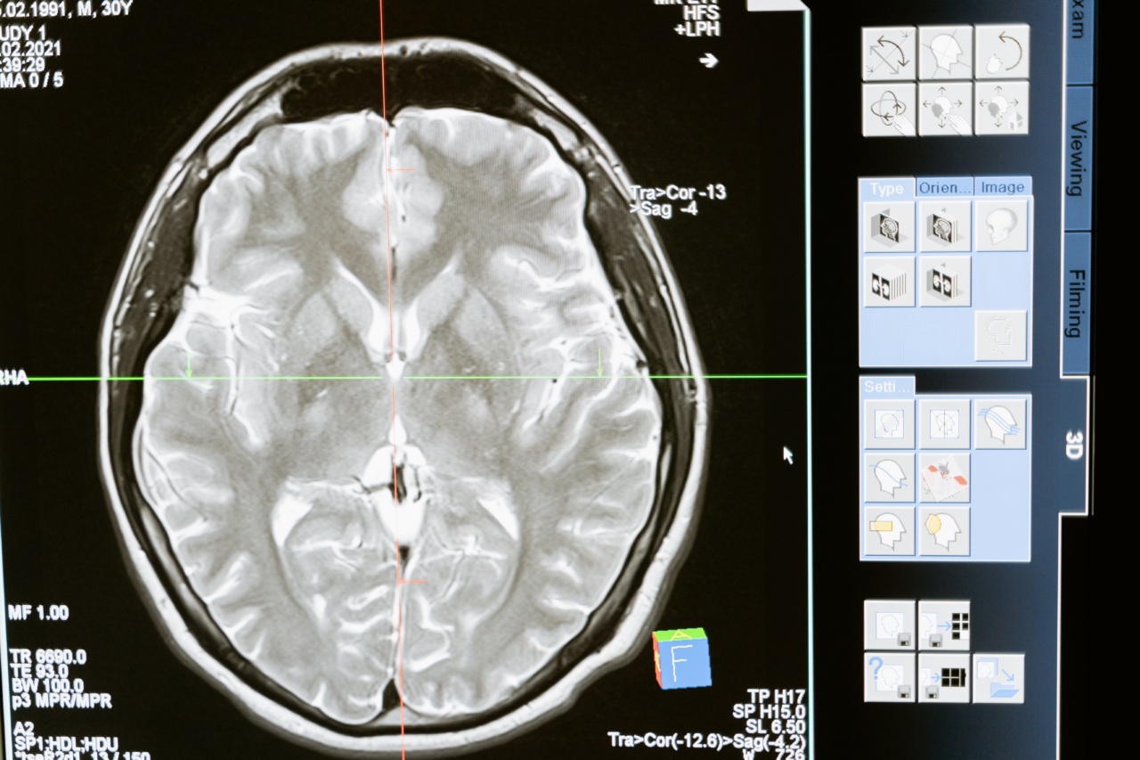
Magnetic Resonance Angiography (MRA)
Magnetic resonance angiography (MRA) is a non-invasive and painless scan that uses a magnetic field and radio waves to produce detailed images of the blood vessels in the brain.
It has been found to be a reliable method of detecting brain hemorrhages, which can help with early diagnosis and prompt treatment.
MRA is a faster and more cost-effective alternative to the traditional CT and MRI scans. It does not require a contrast agent, which makes it safer for patients with kidney problems or allergies to the contrast agent.
Diffusion Tensor Imaging (DTI)
Diffusion tensor imaging (DTI) is a technique that uses magnetic fields and radio waves to measure the movement of water molecules in the brain. This technique is helpful in identifying the extent and severity of brain damage caused by hemorrhage.
DTI has been found to be a reliable method of diagnosing mild to moderate traumatic brain injury and has shown promise in detecting hemorrhages.
Spectroscopy
Spectroscopy is a non-invasive method that uses magnetic fields and radio waves to identify the chemical composition of tissues in the brain. This technique can detect changes in the levels of certain chemicals that are associated with hemorrhages.
Spectroscopy has been found to be useful in detecting hemorrhages in the brainstem, a difficult area to access with other imaging techniques.
Electroencephalography (EEG)
Electroencephalography (EEG) is a non-invasive technique that records the electrical activity of the brain. It has been found to be helpful in identifying brain hemorrhages, especially in newborns.
EEG can detect changes in brain activity that are associated with hemorrhages, such as seizures and abnormal brain wave patterns.
Related Article
Revolutionary smart system detects brain hemorrhage instantly

Computed Tomography (CT) Perfusion Imaging
Computed tomography (CT) perfusion imaging is a technique that uses CT scans to measure the blood flow in the brain. This technique can detect changes in blood flow that are associated with hemorrhages.
CT perfusion imaging is a faster and more cost-effective alternative to traditional CT scans and can help with early diagnosis and prompt treatment of brain hemorrhages.
Magnetic Resonance Imaging (MRI) Perfusion Imaging
Magnetic resonance imaging (MRI) perfusion imaging is a technique that uses MRI scans to measure the blood flow in the brain. This technique can detect changes in blood flow that are associated with hemorrhages.
MRI perfusion imaging is a reliable method of detecting brain hemorrhages and can help with early diagnosis and prompt treatment.
Positron Emission Tomography (PET) Imaging
Positron emission tomography (PET) imaging is a technique that uses a small amount of radioactive material to identify changes in the brain’s activity. This technique can detect changes in brain activity that are associated with hemorrhages.
PET imaging is a useful tool in identifying the location and severity of brain damage caused by hemorrhage and can help with early diagnosis and prompt treatment.
Magnetoencephalography (MEG)
Magnetoencephalography (MEG) is a non-invasive technique that records the magnetic fields generated by the electrical activity in the brain. It has been found to be helpful in identifying the location and extent of brain damage caused by hemorrhage.
MEG can detect changes in brain activity that are associated with hemorrhages, such as abnormal magnetic wave patterns.
Conclusion
The traditional methods of diagnosing brain hemorrhages are time-consuming and expensive. However, advancements in technology have led to the development of faster and more accurate methods of identifying brain hemorrhages.
Magnetic resonance angiography (MRA), diffusion tensor imaging (DTI), spectroscopy, electroencephalography (EEG), computed tomography (CT) perfusion imaging, magnetic resonance imaging (MRI) perfusion imaging, positron emission tomography (PET) imaging, and magnetoencephalography (MEG) are all promising methods of detecting brain hemorrhages.
These techniques can help with early diagnosis and prompt treatment, which can increase the chances of a full recovery and reduce the risk of long-term complications.


