Cardiovascular diseases (CVDs) are the leading cause of mortality worldwide. Early detection and accurate assessment of cardiovascular risk factors play a vital role in preventive care and reducing the burden on healthcare systems.
Computer vision, coupled with artificial intelligence (AI) technology, has emerged as a promising approach to assess cardiovascular risks accurately and efficiently. This article explores the potential of computer vision in cardiovascular risk assessment and discusses its implications in healthcare.
The Role of Computer Vision in Cardiovascular Risk Assessment
Computer vision involves the automated extraction, analysis, and understanding of visual information from images or videos.
In the context of cardiovascular risk assessment, computer vision techniques can be used to analyze medical images, such as cardiac imaging, retinal imaging, or ultrasound scans, to identify relevant biomarkers and risk factors.
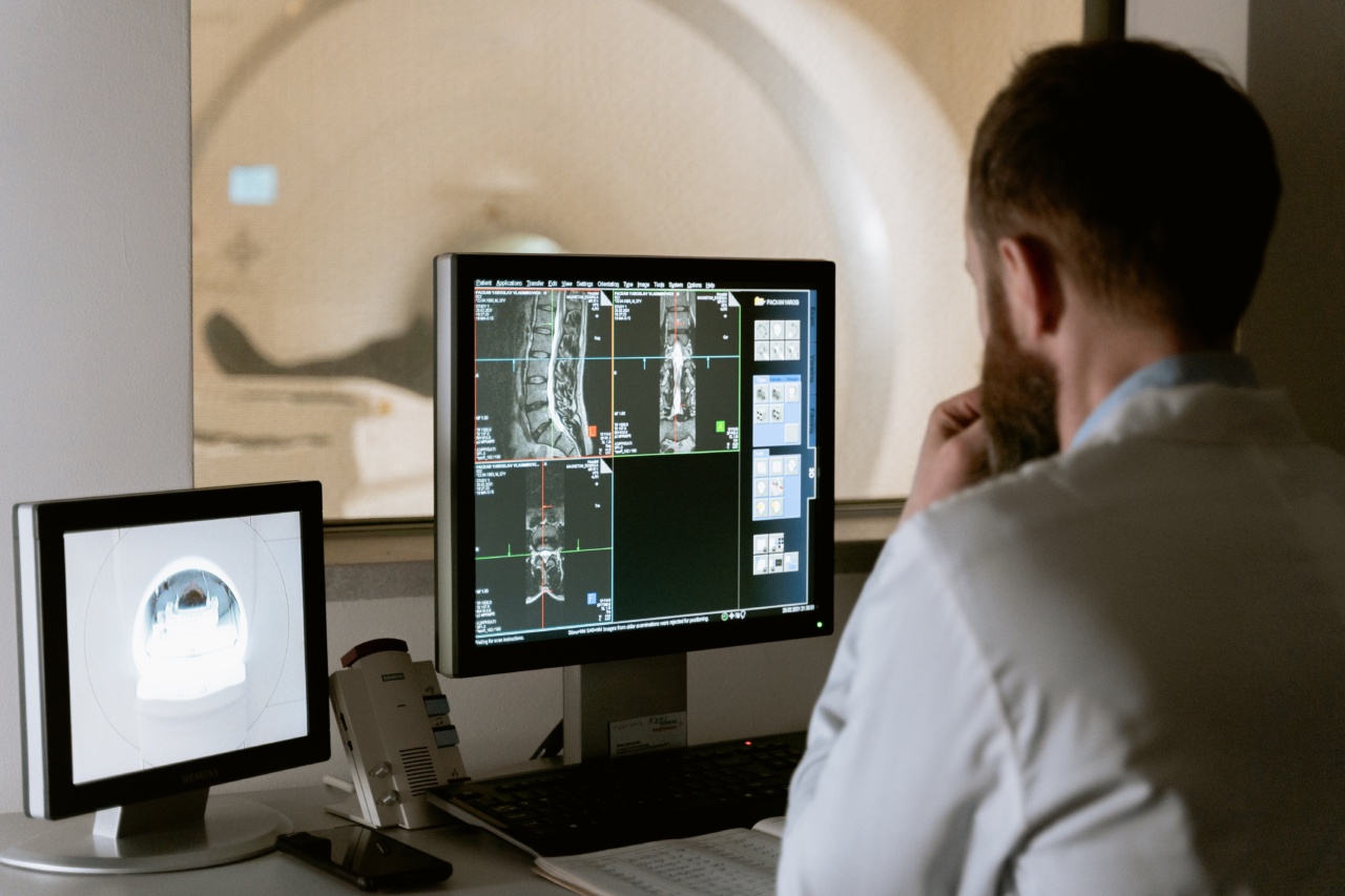
Automated Analysis of Cardiac Imaging
Cardiovascular risk assessment often involves the analysis of cardiac imaging data, such as magnetic resonance imaging (MRI) or computed tomography (CT) scans.
Computer vision algorithms can automatically segment and analyze these images to quantify relevant parameters, such as cardiac chamber volumes, ejection fraction, or myocardial strain. By automating this process, cardiologists can save significant time and effort, while also reducing inter-observer variability.
Retinal Imaging for Cardiovascular Risk Prediction
The retina is the only place in the human body where blood vessels can be directly observed. Research has shown that abnormalities in retinal vasculature are closely linked to cardiovascular diseases.
Computer vision algorithms can analyze retinal images to detect biomarkers, such as arteriovenous ratio, vessel tortuosity, or presence of microaneurysms, which can provide insights into a patient’s cardiovascular risk profile. By leveraging AI technology, these algorithms can learn from large datasets and improve their predictive accuracy over time.
Ultrasound Imaging and Computer Vision
Ultrasound imaging is widely used in cardiovascular diagnostics, allowing medical professionals to visualize the heart and blood flow in real-time.
Related Article
AI System Accurately Predicts Cardiovascular Risk Through Eye Analysis

Computer vision techniques can assist in the automated analysis of ultrasound data, aiding in the identification of cardiac abnormalities and quantification of hemodynamic parameters. AI algorithms can be trained to recognize patterns associated with various cardiovascular conditions, such as hypertrophic cardiomyopathy or valvular diseases, facilitating early detection and intervention.
Machine Learning for Risk Stratification
One of the key advantages of computer vision in cardiovascular risk assessment is its ability to integrate machine learning algorithms.
By training these algorithms on large datasets of medical images and associated clinical data, it is possible to predict the risk of developing CVDs accurately. Machine learning models can take into account various factors, including age, gender, family history, and cardiovascular biomarkers extracted through computer vision techniques, to provide personalized risk assessments and treatment recommendations.
Challenges and Limitations
While computer vision holds great promise in cardiovascular risk assessment, several challenges and limitations need to be addressed. Firstly, the availability of labeled datasets for training machine learning algorithms is crucial.
Data privacy concerns and the need for extensive annotations can hinder the development of robust models. Additionally, the interpretability of computer vision algorithms and their integration into clinical workflows need careful consideration to ensure trust and acceptance among healthcare professionals.
Future Directions and Implications
The ongoing advancements in computer vision and AI technology have the potential to revolutionize cardiovascular risk assessment.
As algorithms become more accurate and reliable, healthcare providers can leverage these tools to enhance their decision-making processes, ultimately leading to better patient outcomes. Furthermore, computer vision can enable remote monitoring and regular assessments by providing automated analysis of imaging data, reducing the need for in-person visits and improving patient convenience.
Conclusion
Computer vision, combined with AI technology, offers a transformative approach to cardiovascular risk assessment.
By automating the analysis of medical images, such as cardiac imaging, retinal imaging, or ultrasound data, computer vision algorithms can provide accurate and efficient risk predictions. While challenges exist, the future of computer vision in cardiovascular care looks promising, and its adoption has the potential to revolutionize preventive healthcare and reduce the burden of cardiovascular diseases.


