Glaucoma is a silent thief of sight, with no early symptoms or warning signs. It is a group of eye conditions that damages the optic nerve, which connects the eye to the brain.
The most common form of glaucoma is called open-angle glaucoma, which is caused by increased pressure inside the eye. This pressure damages the optic nerve over time, leading to irreversible vision loss.
Why Early Detection is Important?
The damage caused by glaucoma is irreversible, and it is impossible to restore vision once it is lost. However, with early detection and treatment, it is possible to slow down the progression of the disease and prevent further vision loss.
Early detection is therefore key to preventing blindness from glaucoma.
Current Methods of Glaucoma Detection
Currently, the main method of detecting glaucoma is by measuring the intraocular pressure (IOP) of the eyes. This is done using a tool called a tonometer, which blows a puff of air onto the surface of the eye to measure the pressure inside.
However, this method is not always accurate, as some people can have normal IOP levels even if they have glaucoma, and vice versa.
Other methods of glaucoma detection include checking the optic nerve for signs of damage, and assessing peripheral vision using a visual field test.
These tests are usually done as part of a comprehensive eye exam, which is recommended for everyone over the age of 40, or for those with a family history of glaucoma.
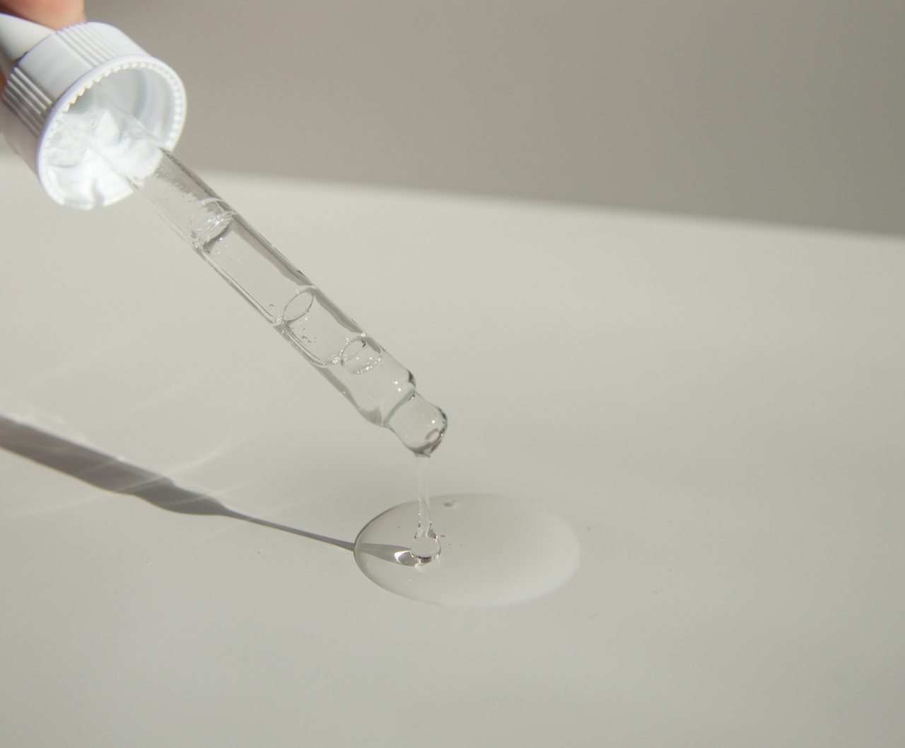
The Tear Test
Recent research has shown that a simple tear test may be a useful tool for detecting glaucoma early.
Related Article
Don’t wait: Early diagnosis of glaucoma requires a tear

The test involves collecting a tear sample from the eye and analyzing it for certain molecules that are known to be involved in the development of glaucoma.
One such molecule is called transforming growth factor beta (TGF-β), which is a protein that promotes the growth of cells and tissues.
In the eye, TGF-β is involved in the development of glaucoma by promoting the activity of certain cells that produce extracellular matrix (ECM), which is a network of proteins that supports the structure of tissues.
In people with glaucoma, the activity of ECM-producing cells is increased, leading to an accumulation of ECM in the trabecular meshwork, which is a network of tissue in the eye that regulates the outflow of aqueous humor, a fluid that helps maintain the shape of the eye. This accumulation of ECM can cause the trabecular meshwork to become clogged, leading to increased IOP levels and damage to the optic nerve.
The tear test works by measuring the levels of TGF-β in the tear sample. If the levels are higher than normal, it could indicate that the patient is at higher risk of developing glaucoma, and further testing may be needed to confirm the diagnosis.
The Benefits of the Tear Test
The tear test has several advantages over current methods of glaucoma detection:.
- It is non-invasive and painless.
- It can be done quickly and easily in a doctor’s office.
- It can provide early detection of glaucoma, before symptoms or vision loss occur.
- It is a sensitive and specific test, meaning that it can accurately detect glaucoma in those who have it, and rule it out in those who do not.
Final Thoughts
The tear test is a promising new tool in the fight against glaucoma. While it is still in the early stages of development, it has the potential to revolutionize the way we detect and treat this debilitating disease.
Early detection is key to preventing vision loss from glaucoma, and the tear test may be just the tool we need to make this a reality.


