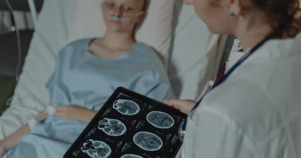
Lung nodules are small growths or lesions that are typically found on the lungs. They can be benign or malignant, and it is crucial to diagnose them accurately to determine the appropriate treatment plan.
Traditionally, lung nodule diagnosis has relied on imaging techniques such as chest X-rays and CT scans. However, with advancements in technology, next-generation tools have emerged that offer more precise and efficient methods for diagnosing lung nodules.
1. Artificial Intelligence (AI) in Lung Nodule Diagnosis
AI has revolutionized various fields, and its potential in lung nodule diagnosis is immense. Machine learning algorithms can analyze large datasets of lung images and identify patterns and characteristics associated with benign or malignant nodules.
These algorithms can then assist radiologists in making accurate diagnoses, reducing human error, and improving patient outcomes.
2. Computer-Aided Detection (CAD) Systems
CAD systems serve as a valuable tool for radiologists in detecting and characterizing lung nodules. These systems use algorithms to analyze medical images and highlight potential abnormalities, aiding radiologists in their diagnostic process.
CAD systems can improve diagnostic accuracy and increase the efficiency of radiologists by acting as a second reader and providing additional insights.
3. Virtual Bronchoscopy
Virtual bronchoscopy is a non-invasive imaging technique that allows the examination of the inside of the airways in the lungs.
It uses advanced computer algorithms to generate three-dimensional (3D) images of the airways, enabling early detection and characterization of lung nodules. Virtual bronchoscopy is a promising tool for diagnosing lung nodules as it avoids the need for invasive procedures.
4. Endobronchial Ultrasound (EBUS)
EBUS is a minimally invasive technique that combines bronchoscopy with ultrasound. It allows for real-time imaging of the airway walls and nearby structures in the lungs.
EBUS can be used to guide biopsies and aspirates of suspicious lung nodules, providing accurate diagnoses with minimal invasiveness. This technology has greatly improved the diagnostic capabilities for lung nodules.
5. Positron Emission Tomography (PET) Scans
PET scans involve the injection of a small amount of radioactive material into the body, which is then detected by a PET scanner. This imaging technique can detect metabolic activity in the body, including in lung nodules.
PET scans are particularly useful in distinguishing between benign and malignant nodules, aiding in treatment decisions and monitoring the effectiveness of treatments.
6. Molecular Profiling
Molecular profiling involves analyzing the genetic and molecular characteristics of lung nodules to determine their subtype and potential response to targeted therapies.
By understanding the genetic makeup of the nodule, physicians can personalize treatment plans and select the most effective medications. Molecular profiling has significantly improved the outcome for patients with lung nodules by enabling personalized medicine.
7. Transbronchial Needle Aspiration (TBNA)
TBNA is a diagnostic procedure that involves using a bronchoscope to obtain tissue samples from lung nodules through a thin needle.
This minimally invasive technique allows for accurate biopsy and diagnosis of nodules that are located within the airways. TBNA has become a standard procedure in the diagnosis of lung nodules, as it provides valuable tissue samples for further analysis and characterization.
8. Liquid Biopsy
Liquid biopsy is a non-invasive method of diagnosing lung nodules by analyzing specific biomarkers in blood samples. It involves testing for circulating tumor cells (CTCs) or tumor DNA fragments in the bloodstream.
Liquid biopsy can provide valuable information about the genetic characteristics of lung nodules, helping physicians select appropriate treatments and monitor treatment response.
9. Virtual Reality (VR) Visualization
VR visualization has recently emerged as a tool for interpreting medical images, including lung nodules. It allows radiologists to immerse themselves in a 3D virtual environment, enabling more detailed and accurate analysis of nodules.
VR visualization enhances the diagnostic process by providing a more intuitive and comprehensive understanding of the spatial relationships within the lungs.
10. Robotic-Assisted Lung Nodule Resection
In cases where surgical intervention is necessary, robotic-assisted procedures have transformed lung nodule resection. Robotic systems provide enhanced precision and dexterity for surgeons, allowing for minimally invasive removal of nodules.
These systems offer improved visualization, precise instrument control, and reduced patient trauma, leading to faster recovery times and improved patient outcomes.


