Prostate cancer is a prevalent form of cancer that affects men worldwide. Detecting prostate cancer early is essential to increase the chances of successful treatment.
However, early prostate cancer can often display no symptoms making it challenging to diagnose until it has progressed. That’s why many men choose to undergo regular screening exams to check for any signs of prostate cancer.
Common Prostate Cancer Screening Examinations
The most common prostate cancer screening exams are the prostate-specific antigen (PSA) blood test and the digital rectal exam (DRE).
Prostate-Specific Antigen Blood Test
The PSA test measures the levels of PSA in a man’s blood. PSA is a protein that the prostate gland produces, and elevated PSA levels can indicate the presence of prostate cancer.
High PSA levels, however, do not definitively diagnose prostate cancer. It’s essential to note that other conditions, such as an enlarged or inflamed prostate, can increase PSA levels.
Digital Rectal Exam
The DRE involves a healthcare provider using a gloved finger to feel the prostate through the rectum. Abnormalities, such as lumps, firmness, or enlargement, may indicate prostate cancer.
DRE is a useful tool for highlighting the presence of physical lumps or abnormalities within the prostate gland.
The Limitations of Common Prostate Cancer Screening Tests
While both PSA and DRE are prevalent prostate cancer screening methods, they have limitations. .
Limitations of the PSA Test
PSA test results can provide false-positive results, meaning elevated PSA levels are not due to cancer. False-negative results are also possible, with PSA levels displaying within the normal range even when prostate cancer is present.
In addition, PSA levels can vary naturally, making it challenging to interpret results accurately. .
Limitations of the Digital Rectal Exam
The DRE has limitations in detecting prostate cancer in the gland’s outer regions. As such, it can often miss the presence of prostate cancer unless the disease is more advanced.
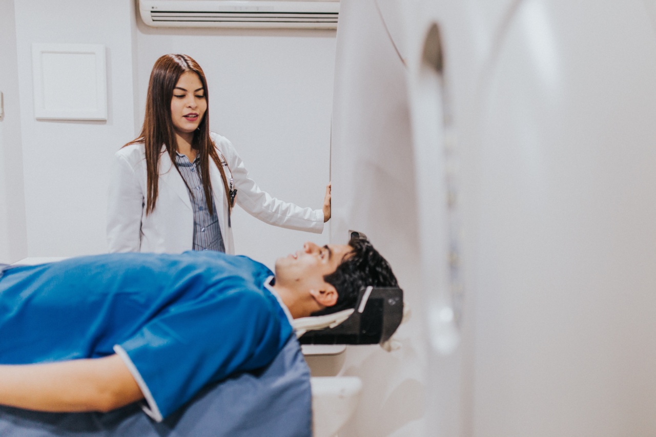
MRI-Guided Biopsy: An Alternative Prostate Cancer Diagnosis
MRI-guided biopsy provides an accurate prostate cancer diagnosis with a high degree of accuracy.
Rather than just screening for cancer, an MRI-guided biopsy takes targeted tissue samples from the prostate gland, reducing the risk of missing some cancers. It also reduces the possibility of patients undergoing unnecessary biopsies, which carry their risks.
What is an MRI-guided Biopsy?
An MRI-guided biopsy is a prostate cancer diagnostic procedure that uses a magnetic resonance imaging (MRI) scanner to guide a needle through the rectum and towards the prostate gland.
The ensuing needle samples small tissue pieces from the areas of the gland where an MRI indicates a biopsy is necessary or required. The samples are then analyzed at a laboratory for the presence of cancerous cells.
What are the benefits of an MRI-guided biopsy?
Some of the benefits of an MRI-guided biopsy include:.
Related Article
A Foolproof Prostate Cancer Diagnostic Test

Increased accuracy
An MRI-guided biopsy can pinpoint the location of the cancerous cells within the prostate gland.
It detects better areas of high-grade disease and reveals the disease’s extent better than other diagnostic procedures, such as a DRE or PSA blood test, for example.
Minimal invasiveness
Unlike other prostate cancer biopsies, MRI-guided biopsies are minimally invasive. The procedure doesn’t require general anesthesia, reducing the risks and discomfort usually associated with more invasive surgical biopsies.
Reduced risk of complications
The risks of complications, such as prostate infection and bleeding, are lower with MRI-guided biopsies than with other more traditional methods. .
Precise biopsy placement
With MRI-guided biopsies, the healthcare professional performing the biopsy can visualize the precise location of a patient’s tumor. This allows them to access hard-to-reach areas of the gland, increasing the chances of an accurate diagnosis.
Who Needs an MRI-guided Biopsy?
For men with elevated PSA levels, irregular DRE results, or other factors that suggest a higher possibility of developing prostate cancer, an MRI-guided biopsy may be beneficial.
It can also be used in men whose previous biopsies have been negative but are still experiencing symptoms associated with prostate cancer.
The Procedure for an MRI-guided Biopsy
The MRI-guided biopsy procedure involves several steps:.
MRI scan
The first step involves a high-resolution MRI scan to create images of the prostate gland. The images help the healthcare professional identify any suspicious areas within the gland and determine where the biopsy needs to be directed.
Under ideal circumstances, the MRI scan is of high-quality and is performed by a highly experienced technician to create accurate images.
Biopsy planning
Using the MRI scan images, the healthcare professional identifies the number and location of biopsies required. The results help them to plan the biopsy and determine the number of needle samples required to achieve a reliable diagnosis.
Biopsy procedure
The biopsy procedure involves a local anesthetic to numb the area and a biopsy needle advances under MRI guidance towards the prostate gland. Once correctly positioned, the needle takes small tissue samples from the gland.
The procedure is relatively painless, and few men require pain control medication once the biopsy is complete.
Laboratory analysis
The biopsy samples are then analyzed at a laboratory to determine whether any cancerous cells are present in the tissue samples.
Conclusion
Early prostate cancer detection is essential for successful treatment. However, common prostate cancer screening exams, such as the PSA blood test and DRE, can often provide inaccurate results.
MRI-guided biopsy, on the other hand, is a highly accurate diagnostic tool that enables healthcare professionals to remove small tissue samples precisely and diagnose prostate cancer with a high degree of accuracy. If you suspect you have prostate cancer or have concerns about prostate cancer, talk to your healthcare provider for guidance and support in getting screened.


