MRI (Magnetic Resonance Imaging) scanning is an advanced medical imaging technique that provides detailed and clear images of the various parts of the body. It utilizes strong magnetic fields and radio waves to generate these images.
MRI scanning has revolutionized the field of diagnostic medicine, enabling physicians to accurately diagnose and devise treatment plans for various medical conditions. In this article, we will delve into the basics of MRI scanning, including how it works, its applications, and its advantages over other imaging techniques.
How does MRI scanning work?
MRI scanning involves a complex interplay of magnetic fields and radio waves. Firstly, the patient is positioned inside a large and cylindrical MRI machine. This machine consists of a strong magnet that creates a powerful and uniform magnetic field.
When the patient enters the magnetic field, the protons in their body align themselves with this field.
Next, radio waves are emitted into the body. These radio waves have a specific frequency and energy that is absorbed by the aligned protons.
When the radio waves are turned off, the protons release the absorbed energy in the form of electromagnetic signals. These signals are then picked up by special receivers in the MRI machine.
The receivers in the machine detect the signals and convert them into detailed images using sophisticated computer software. These images can then be interpreted by radiologists to identify any abnormalities or areas of concern.
Advantages of MRI scanning
MRI scanning offers several advantages over other imaging techniques such as X-rays or CT scans. Firstly, it does not involve the use of ionizing radiation, making it a safer option for patients, especially those who require repeated imaging studies.
This makes MRI scanning particularly suitable for young children, pregnant women, and individuals with sensitivity or allergies to contrast agents.
Furthermore, MRI scans provide highly detailed images that are superior in clarity and contrast compared to other imaging techniques.
This allows radiologists and physicians to accurately assess various structures within the body, including soft tissues, organs, joints, and blood vessels. MRI is particularly valuable for evaluating brain and spinal cord conditions, tumors, joint injuries, and cardiovascular diseases.
MRI scanning also allows for imaging in multiple planes, by adjusting the orientation of the imaging slices.
This provides comprehensive and multi-dimensional views of the structures being evaluated, aiding in efficient diagnosis and treatment planning.
Preparing for an MRI scan
Before undergoing an MRI scan, certain preparations may be required. The patient will be asked to remove any metallic objects such as jewelry, watches, belts, or hairpins, as these can interfere with the magnetic field of the machine.
Additionally, individuals with certain metallic implants or devices, such as pacemakers or cochlear implants, may not be eligible for MRI scanning due to possible risks and interference.
Patients will usually be instructed to wear a gown provided by the imaging facility.
It is important to inform the healthcare provider about any pre-existing medical conditions, especially those related to kidney function, as some contrast agents used in MRI scans can affect the kidneys.
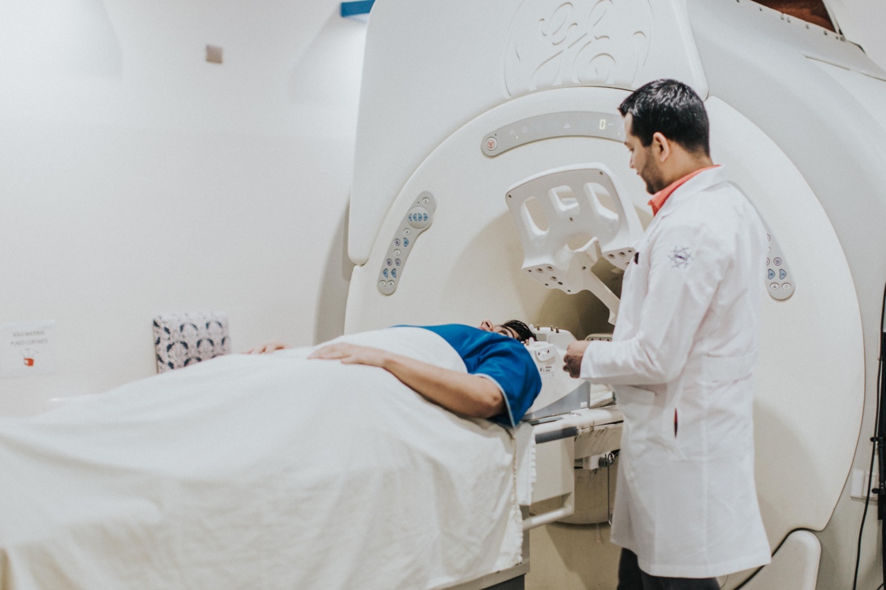
The MRI scanning procedure
During the MRI scanning procedure, the patient will be positioned on a movable bed that slides into the cylindrical MRI machine. It is important to lie still during the scan to avoid blurring the images.
Related Article
The science behind magnetic resonance imaging

Any movement can lead to distortions in the final images, reducing their diagnostic value.
Some MRI scans might require the use of a contrast agent. This is usually injected intravenously to improve the visibility of certain structures or highlight areas of concern.
The contrast agent used in MRI scans is typically gadolinium-based, and is generally well tolerated by most patients. However, individuals with a history of allergic reactions or kidney problems should inform their healthcare provider prior to the scan.
The MRI scanning process can take anywhere from a few minutes to over an hour, depending on the area being imaged and the complexity of the study.
During the scan, the patient will hear loud knocking or thumping noises, which are created by the magnetic field and radio wave pulses. Earplugs or headphones are usually provided to minimize discomfort and protect hearing.
Types of MRI scans
There are various types of MRI scans that are used to visualize different parts of the body. Some common types include:.
1. Brain MRI
A brain MRI is used to examine the neurological structures and detect abnormalities such as tumors, hemorrhages, or signs of stroke. It is a non-invasive procedure that offers detailed visualization of the brain and its different regions.
2. Spine MRI
A spine MRI is commonly used to evaluate conditions affecting the spinal cord, vertebrae, or intervertebral discs. It helps in diagnosing spinal cord injuries, herniated discs, stenosis, or infections.
3. Abdominal MRI
An abdominal MRI is used to analyze the structures and organs within the abdominal region, including the liver, kidneys, pancreas, and intestines. It can help identify tumors, cysts, or abnormalities in these organs.
4. Joint MRI
A joint MRI is performed to assess the bones, ligaments, tendons, and cartilage of the joints. It is commonly used for diagnosing injuries or diseases affecting the knees, shoulders, wrists, or hips.
Risks and considerations
While MRI scanning is generally considered safe, there are certain risks and considerations to be aware of.
The strong magnetic field of the MRI machine can attract metallic objects in the vicinity, causing serious injuries or damage if they are not properly removed. It is imperative to ensure that no metallic objects are present during the scan.
Some individuals may experience claustrophobia or anxiety during the MRI scan, as the cylindrical machine can be confining.
However, open MRI machines, which are less enclosed, are now available and can be a suitable alternative for claustrophobic individuals.
As mentioned earlier, contrast agents are sometimes used during MRI scans. While adverse reactions to these agents are rare, they can occur.
Allergies, especially to gadolinium-based agents, should be discussed with the healthcare provider prior to the scan. In addition, individuals with compromised kidney function should exercise caution, as the contrast agent can have a negative impact on the kidneys.
Conclusion
MRI scanning is a powerful diagnostic tool that allows for detailed and accurate imaging of the body’s structures. It provides valuable information to physicians for diagnosing and treating various medical conditions.
With its numerous advantages, including safety, clarity, and multi-dimensional views, MRI scanning has become an indispensable part of modern healthcare. By understanding the basics of this imaging technique, patients can make informed decisions and feel more confident during the scanning process.


