The human eye is a fascinating and complex structure that is responsible for our sense of sight. It serves as a window through which we perceive the world and provides valuable information about our environment.
Advancements in medical technology have made it possible to analyze the structures and processes of the eye in greater detail.
Scientists and researchers can now use advanced imaging techniques to capture images of the eye’s interior, allowing them to study the subtle changes and abnormalities that may lead to vision problems or eye diseases.
What is the Pupil?
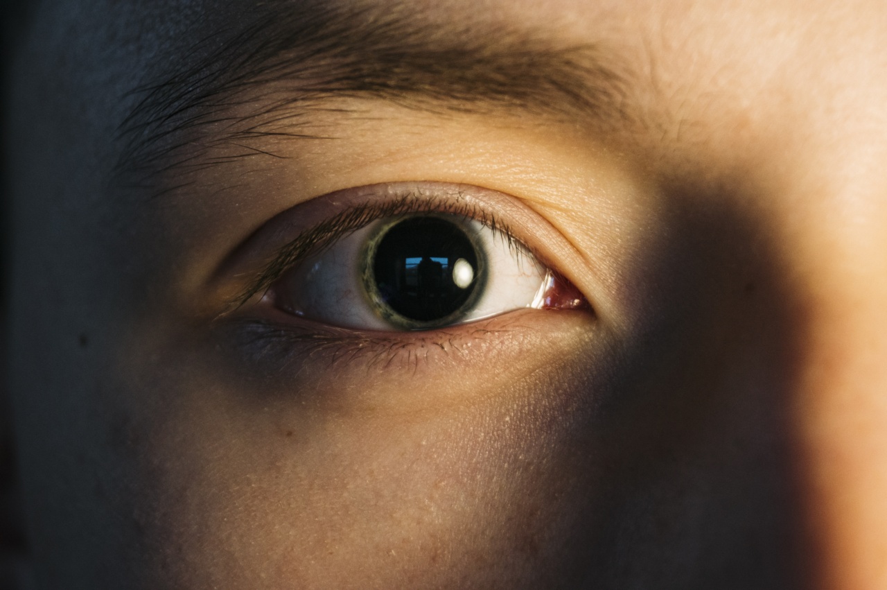
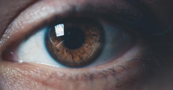
The pupil is the black circular opening in the center of the iris, the colored part of the eye. It regulates the amount of light that enters the eye, expanding or contracting as needed to adjust to the levels of light in the environment.
Dilation of the pupil is a common technique used to examine the interior of the eye. It involves the use of eye drops to widen the pupil, allowing doctors a better view of the internal structures.
However, this process can be uncomfortable and inconvenient for patients, especially when it affects their ability to see clearly afterward.
The Innovation in Imaging Technique
A new imaging technique developed by scientists at the University of Illinois at Urbana-Champaign uses optical coherence tomography (OCT) to capture detailed, three-dimensional images of the eye’s interior without dilation of the pupil.
OCT uses light waves to create high-resolution images of the interior of the eye. The technology sends light waves into the eye, where they are reflected back and recorded by a computer.
The software then creates a detailed image of the structures within the eye, including the retina, optic nerve, and macula.
The new technique, called Angio-Oct, combines OCT with an additional imaging system that captures images of the blood vessels within the eye.
This provides doctors with even more information about the health of the eye and allows them to detect early signs of disease.
The Benefits of No-Pupil-Dilation Imaging
The benefits of this new imaging technique are clear. Since there is no need for pupil dilation, patients can avoid the discomfort and inconvenience associated with the procedure.
They can return to their daily routines immediately after the examination without experiencing blurred vision or sensitivity to light.
Additionally, the images captured by the Angio-Oct technique provide doctors with a clear and detailed view of the structures and blood vessels within the eye.
This allows them to detect even the slightest changes in the eye’s health and diagnose eye diseases at an early stage when they are easiest to treat.
The Medical Applications of Angio-Oct
The Angio-Oct imaging technique has a wide range of medical applications.
It can be used to diagnose and monitor diseases such as age-related macular degeneration, diabetic retinopathy, and glaucoma, which are major causes of vision loss and blindness worldwide.
It can also be used to study the effects of diseases, such as multiple sclerosis, on the health of the eye.
Researchers can use the technique to analyze how changes in the eye’s blood vessels reflect changes in the rest of the body, providing valuable insight into the progression of the disease.
The advances made in imaging techniques such as Angio-Oct have revolutionized our ability to examine and diagnose eye diseases.
They provide doctors with a powerful tool to monitor the health of the eye and offer early intervention for diseases that can cause permanent vision loss.
The Future of Eye Imaging Technology
The Angio-Oct imaging technique represents a significant advancement in the field of eye imaging technology.
It provides doctors with a clear and detailed view of the eye’s interior without the need for uncomfortable procedures such as pupil dilation.
As research and development continue, we can expect further innovations in eye imaging technology that will provide even more comprehensive views of the eye’s interior.
These technologies will be vital in the diagnosis and treatment of eye diseases, helping to preserve the vision of millions of people worldwide.
Conclusion
The development of the Angio-Oct imaging technique marks a significant milestone in the field of eye imaging technology.
It allows doctors to capture detailed three-dimensional images of the eye’s interior without the need for pupil dilation, making it a more comfortable and less invasive procedure for patients.
The benefits of this new imaging technique are clear. It provides doctors with a powerful tool to diagnose and monitor diseases, offers early intervention for eye diseases, and helps to preserve the vision of millions of people worldwide.


