Cervical cancer is a type of cancer that develops in the cervix, which is the lower part of the uterus. It is usually caused by the human papillomavirus (HPV) and can be prevented through vaccination and regular screening.
In this article, we will discuss advanced screening methods for cervical cancer.
1. Liquid-Based Cytology (LBC)
Liquid-based cytology (LBC) is a method of cervical cancer screening in which a sample of cells is collected from the cervix and placed in a liquid medium. The cells are then examined under a microscope to look for abnormalities.
LBC has several advantages over traditional Pap smears, including:.
- Higher accuracy
- Reduced false-negative results
- Ability to test for HPV at the same time
- Improved sample preservation
2. Human Papillomavirus (HPV) Testing
HPV testing is a method of cervical cancer screening that looks for the presence of the human papillomavirus (HPV) in cervical cells. HPV is the primary cause of cervical cancer and is responsible for over 99% of cases.
HPV testing can be done on its own or in conjunction with other screening methods, such as Pap smears or LBC. HPV testing is recommended for women over the age of 30.
3. Visual Inspection with Acetic Acid (VIA)
Visual inspection with acetic acid (VIA) is a simple and low-cost method of cervical cancer screening that involves applying a solution of acetic acid to the cervix and examining it for abnormalities with the naked eye or a magnifying device.
VIA is particularly useful in low-resource settings where more advanced screening methods may not be available.
4. Visual Inspection with Lugol’s Iodine (VILI)
Visual inspection with Lugol’s iodine (VILI) is another low-cost method of cervical cancer screening that involves applying a solution of Lugol’s iodine to the cervix and examining it for abnormalities.
VILI is particularly useful for detecting precancerous lesions and is often used in conjunction with other screening methods.
5. Colposcopy
Colposcopy is a diagnostic procedure in which a medical professional examines the cervix using a colposcope, a specialized instrument that uses a light and a magnifying lens to look for abnormalities.
During the colposcopy, the medical professional may take a biopsy, which involves removing a small sample of tissue from the cervix for further testing.
6. Cone Biopsy
A cone biopsy is a surgical procedure in which a cone-shaped piece of tissue is removed from the cervix for further testing. Cone biopsies are usually done when abnormal cells are found during a Pap smear or other screening method.
The tissue removed during a cone biopsy can be examined under a microscope to determine if the cells are cancerous or precancerous.
7. Magnetic Resonance Imaging (MRI)
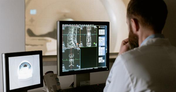
Magnetic resonance imaging (MRI) is a non-invasive diagnostic imaging tool that uses a magnetic field and radio waves to create detailed images of the body.
MRI can be used to detect and stage cervical cancer, as well as to monitor the progress of treatment.
8. Computed Tomography (CT) Scan
Computed tomography (CT) is a diagnostic imaging tool that uses x-rays and computer technology to create detailed images of the body. CT scans can be used to detect and stage cervical cancer, as well as to monitor the progress of treatment.
9. Positron Emission Tomography (PET) Scan
Positron emission tomography (PET) is a diagnostic imaging tool that uses a small amount of radioactive material to create three-dimensional images of the body.
PET scans can be used to detect and stage cervical cancer, as well as to monitor the progress of treatment.
10. Sentinel Lymph Node Biopsy
A sentinel lymph node biopsy is a surgical procedure in which the sentinel lymph node, which is the first lymph node to which cancer is likely to spread, is removed and examined for the presence of cancer cells.
Sentinel lymph node biopsy can be used to determine the stage of cervical cancer and to guide treatment decisions.


