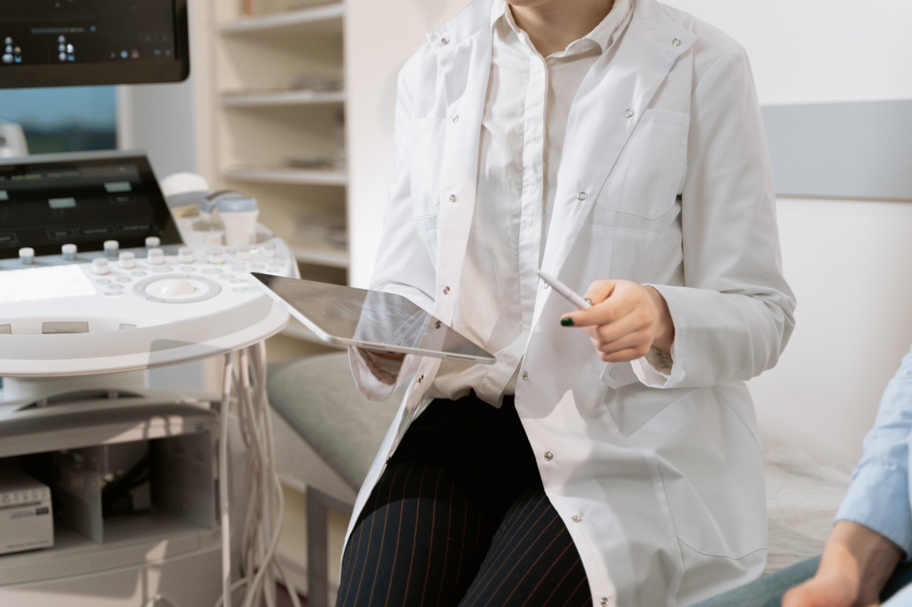
Diagnostic breast ultrasound is a noninvasive and relatively low-cost imaging technique that uses high-frequency sound waves to produce images of the breast tissue.
It is a widely used diagnostic tool to assist in the detection and diagnosis of breast cancer. In recent years, advancements in diagnostic breast ultrasound have improved the accuracy and efficiency of breast cancer diagnosis and treatment. This article explores some of these advancements.
The Advancements
Automated Breast Ultrasound
Automated Breast Ultrasound (ABUS), also known as Automated Whole Breast Ultrasound (AWBU), is a recent advancement in breast ultrasound technology.
ABUS incorporates a computer system that takes and analyzes multiple images of the entire breast, providing a three-dimensional view of the breast tissue. This technology increases the sensitivity of breast cancer detection and has been proven to be effective in detecting breast cancer in women with dense breast tissue.
Elastography
Elastography is a diagnostic technique that uses ultrasound to measure tissue elasticity. This technique has been used to identify cancerous breast lesions that are stiff, which is a characteristic of cancerous tissue.
The use of elastography has improved the accuracy of breast cancer diagnosis by distinguishing between benign and malignant breast lesions.
Contrast-Enhanced Ultrasound
Contrast-enhanced ultrasound (CEUS) is a technique that uses a contrast agent to improve the visualization of blood vessels and tissue perfusion. This technique has been used to assess breast lesions by providing information about blood flow patterns.
In cases where a biopsy is required, CEUS can help guide the biopsy needle to the correct location. CEUS has shown to be useful in the evaluation of suspicious breast lesions especially in patients who are unable to undergo MRI.
Shear Wave Elastography
Shear Wave Elastography (SWE) is an advanced form of elastography, which measures the stiffness of tissue using shear waves. Shear waves are mechanical waves that propagate through tissue and produce an image of tissue stiffness.
SWE can be used to identify the tissue stiffness of breast lesions, which can help distinguish between benign and malignant lesions. SWE has been shown to be more accurate than conventional elastography and improves the accuracy of breast cancer diagnosis.
Molecular Breast Imaging
Molecular Breast Imaging (MBI) is a diagnostic technique that uses small amounts of radioactive material to create images of the breast tissue. This technique is used to detect cancerous lesions in dense breast tissue.
MBI has shown to be more effective than mammography in detecting small breast tumors, especially in women with dense breast tissue.
Automated Lesion Detection
Automated lesion detection uses computer algorithms to search for abnormalities in breast images. The system analyzes the images for suspicious lesions, which are then prioritized based on the likelihood of malignancy.
Related Article
Groundbreaking Technologies in Breast Ultrasound

This method has been shown to be more efficient and accurate than manual detection, reducing the number of false-positive results.
Artificial Intelligence
Artificial Intelligence (AI) is a rapidly developing field that has found uses in many different areas of medicine, including diagnostic breast ultrasound.
AI algorithms can help identify and classify suspicious breast lesions, assisting radiologists in the diagnosis of breast cancer. AI technology has shown to increase the accuracy of breast cancer diagnosis and has the potential to reduce the workload for radiologists.
Telemedicine
Telemedicine allows healthcare professionals and patients to communicate remotely, using technology such as video conferencing.
Telemedicine has been used in diagnostic breast ultrasound to provide remote consultations and eliminate the need for patients to travel to a healthcare facility. This technology has the potential to improve access to healthcare in remote areas and increase the efficiency of breast cancer diagnosis and treatment.
Portable Ultrasound
Portable Ultrasound is a technology used to provide remote ultrasounds in mobile settings. This technology can be used to diagnose breast cancer in remote areas and in emergency situations.
Portable ultrasound machines can range from small handheld devices to larger, more complex machines, allowing for flexibility in use.
3D Printing
3D Printing is a rapidly developing technology that has found use in many different areas of medicine.
In diagnostic breast ultrasound, 3D printing is used to create physical models of breast tumors, which can be used to improve patient communication and education. It can also be used to assess the size and location of a breast tumor, assisting in the planning of surgical procedures.
Conclusion
The advancements in diagnostic breast ultrasound have improved our ability to detect and diagnose breast cancer.
The use of technology such as automated breast ultrasound, elastography, and contrast-enhanced ultrasound has improved the accuracy and efficiency of breast cancer diagnosis. Artifical intelligence and telemedicine have the potential to improve access to healthcare and reduce the workload for radiologists.
Portable ultrasound and 3D printing have the potential to improve patient communication and education, and assist in surgical planning. These advancements are transforming the field of diagnostic breast ultrasound and improving breast cancer diagnosis and treatment.


