Depression is a prevalent mental health disorder that affects millions of people worldwide. It is characterized by persistent feelings of sadness, loss of interest or pleasure, and a range of physical and cognitive symptoms.
While the exact cause of depression remains unknown, researchers have made significant progress in understanding the biological basis of the disorder through brain imaging studies. In this article, we will explore what brain imaging has revealed about depression and its potential implications for diagnosis and treatment.
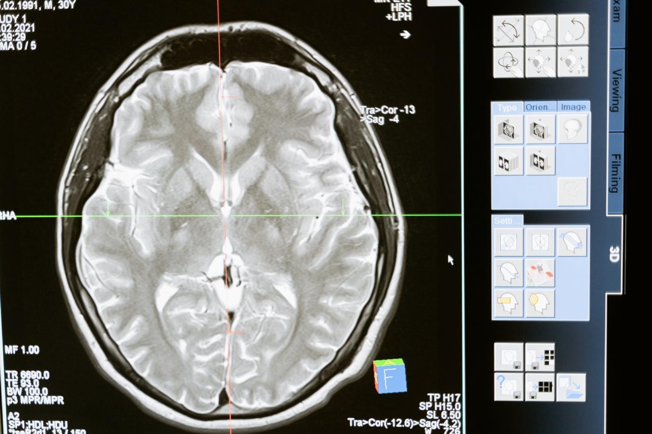
Understanding Brain Imaging
Brain imaging techniques allow scientists to visualize and study the structure and function of the brain.
There are various methods used for brain imaging, including magnetic resonance imaging (MRI), positron emission tomography (PET), functional MRI (fMRI), and electroencephalography (EEG). Each method provides unique insights into different aspects of brain activity and connectivity.
Neuroanatomical Findings in Depression
Numerous studies using MRI have revealed structural differences in the brains of individuals with depression compared to those without the condition.
These differences include reduced volume in certain brain regions, particularly the hippocampus, amygdala, and prefrontal cortex. Additionally, abnormalities in the white matter pathways connecting different brain regions have been observed in depressed individuals.
Neurochemical Imbalances
Brain imaging studies have also shed light on the role of neurotransmitters in depression. Neurotransmitters are chemical messengers that facilitate communication between brain cells.
Imbalances in neurotransmitter levels, particularly serotonin, norepinephrine, and dopamine, have been implicated in depression. PET scans have shown altered receptor binding and availability of these neurotransmitters in individuals with depression.
Functional Connectivity Alterations
Functional connectivity refers to the synchronized activity between different brain regions. Resting-state fMRI studies have highlighted disrupted functional connectivity patterns in individuals with depression.
Specifically, decreased connectivity within the default mode network, involved in self-referential thinking, and increased connectivity within the amygdala and other emotion-regulation circuits have been observed. These alterations may underlie the emotional and cognitive symptoms commonly experienced in depression.
Stress and Neuroplasticity
Chronic stress plays a significant role in the development and maintenance of depression. Recent research using MRI has shown that stress can lead to structural changes in the brain, particularly in the hippocampus.
Related Article
Depression and the Brain: An Overview

The hippocampus is involved in memory, learning, and emotion regulation. Persistent stress may impair the functioning of the hippocampus, contributing to the symptoms of depression. Furthermore, neuroplasticity, the brain’s ability to adapt and reorganize, is also believed to be impacted by stress and depression.
Predictive Value of Brain Imaging
While brain imaging techniques have provided valuable insights into the neurobiological aspects of depression, they have not yet been translated into routine clinical use for diagnosis or treatment.
However, researchers have begun exploring the potential of using brain imaging as a predictive tool. For example, machine learning algorithms have been trained to predict treatment response in depressed individuals based on their brain imaging data.
This field, known as psychiatric neuroimaging, holds promise for personalized treatment approaches in the future.
Limitations and Future Directions
Despite the advancements in brain imaging studies of depression, there are several challenges and limitations that need to be addressed.
Firstly, the heterogeneity of depression as a disorder makes it difficult to identify consistent biomarkers across all individuals. Moreover, the majority of studies have focused on adults, limiting our understanding of depression in children and adolescents.
Future research should aim to overcome these limitations and explore the potential of brain imaging as a tool for early diagnosis, prevention, and personalized treatment of depression.
Conclusion
Brain imaging studies have significantly enhanced our understanding of the neurobiological underpinnings of depression.
Structural, functional, and neurochemical alterations have been observed in individuals with depression, providing insights into the mechanisms of this complex disorder. While brain imaging is not currently used for routine diagnosis or treatment of depression, ongoing research holds promise for its future clinical utility.
By unraveling the mysteries of the brain, we can develop more targeted and effective interventions for individuals suffering from depression.


