According to the World Health Organization (WHO), stroke is the second leading cause of death globally, responsible for approximately 11% of all deaths.
It is estimated that 15 million people suffer from stroke each year, with survivors often experiencing long-term disabilities. Early detection and prevention of strokes are therefore crucial in saving lives and reducing the burden on healthcare systems.
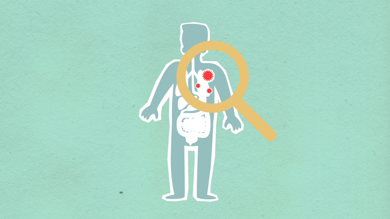
In recent years, a breakthrough examination has emerged that helps predict stroke risk and occurrence, offering hope for better stroke prevention and treatment.
Understanding Stroke: Causes and Risk Factors
Before delving into the breakthrough examination, it is important to understand the basics of stroke.
Stroke occurs when there is a disruption of blood flow to the brain, either due to a blockage in the blood vessels (ischemic stroke) or a rupture of blood vessels (hemorrhagic stroke). The brain cells are deprived of oxygen and nutrients, leading to their death within minutes.
The severity of the stroke and its long-term effects depend on various factors, including the area of the brain affected and the promptness of medical intervention.
There are several risk factors that increase the likelihood of stroke, some of which are modifiable while others are not. Non-modifiable risk factors include age, gender, race, and family history of stroke.
Modifiable risk factors, on the other hand, can be controlled through lifestyle changes or medical interventions. These include high blood pressure, smoking, obesity, diabetes, high cholesterol levels, sedentary lifestyle, and excessive alcohol consumption.
The Breakthrough Examination: How Does It Work?
The breakthrough examination that helps predict stroke risk and occurrence is called carotid artery ultrasound.
It is a non-invasive imaging technique that uses sound waves to produce real-time images of the carotid arteries, the major blood vessels located in the neck that supply blood to the brain. This examination allows healthcare professionals to assess the presence of plaque buildup in these arteries.
Plaque buildup, also known as atherosclerosis, is a condition characterized by the formation of fatty deposits and other substances on the inner walls of the arteries.
Over time, these deposits can harden, narrow the arterial lumen, and disrupt blood flow. In the carotid arteries, plaque buildup can significantly increase the risk of stroke, as it can lead to the detachment of plaque fragments or the formation of blood clots that can travel to the brain.
During a carotid artery ultrasound, a handheld device called a transducer is passed over the neck area. The transducer emits high-frequency sound waves, which bounce off the tissues and blood vessels and are picked up by the transducer again.
The data collected by the transducer is processed by a computer to generate real-time images that can be displayed on a screen. These images allow healthcare professionals to visualize the carotid arteries and identify any abnormalities or plaque buildup.
Benefits of Carotid Artery Ultrasound in Stroke Prediction
The carotid artery ultrasound examination offers several benefits in predicting stroke risk and occurrence, making it a breakthrough tool in stroke prevention and management. Some of the key advantages of this examination are:.
1. Early Detection of Plaque Buildup
Carotid artery ultrasound enables the early detection of plaque buildup in the carotid arteries. By identifying the presence and extent of plaque, healthcare professionals can assess the severity of atherosclerosis and determine the risk of stroke.
Early detection allows for timely interventions and lifestyle modifications to prevent the progression of plaque buildup and reduce the risk of stroke.
Related Article
New examination method predicts stroke and stroke risk

2. Identification of High-Risk Individuals
The carotid artery ultrasound examination helps identify individuals who are at high risk of stroke due to significant plaque buildup in their carotid arteries.
By identifying high-risk individuals, healthcare professionals can prioritize preventive measures and interventions, such as medication management, blood pressure control, and cholesterol-lowering strategies, to reduce the risk of stroke and its potential complications.
3. Personalized Treatment Planning
Each individual’s risk of stroke is unique, and treatment strategies need to be tailored accordingly.
Carotid artery ultrasound provides essential information on the extent and location of plaque buildup, allowing healthcare professionals to develop personalized treatment plans. For example, individuals with severe plaque buildup may require surgical interventions, such as carotid endarterectomy or stenting, to restore adequate blood flow and reduce the risk of stroke.
4. Monitoring Treatment Effectiveness
After initiating preventive measures or treatment interventions, it is essential to monitor their effectiveness in reducing plaque buildup and preventing stroke.
Carotid artery ultrasound can be used as a follow-up tool to assess the progression or regression of plaque and to evaluate the response to treatment. This enables healthcare professionals to make necessary adjustments in the management plan to optimize outcomes.
5. Minimally Invasive and Safe
Carotid artery ultrasound is a non-invasive examination that is generally safe and well-tolerated by patients. Unlike invasive procedures, such as angiography, it does not require the insertion of catheters or the use of contrast agents.
The absence of radiation exposure also adds to the safety profile of this examination. Therefore, it can be routinely used in both outpatient and inpatient settings without significant risks or discomfort for the patients.
Future Directions and Limitations
While carotid artery ultrasound holds great promise in stroke prediction, there are certain limitations and areas for further research.
Firstly, this examination primarily focuses on the assessment of carotid artery plaques and may not provide a comprehensive evaluation of other potential causes of stroke. Other imaging techniques, such as magnetic resonance imaging (MRI) or computed tomography angiography (CTA), may be needed in some cases for a more thorough evaluation.
Secondly, the interpretation of carotid artery ultrasound images requires expertise and experience.
Healthcare professionals who perform and interpret these examinations need to have the necessary training to accurately identify and assess plaque buildup. Initiatives to standardize training and certification in carotid artery ultrasound interpretation are essential to ensure consistent and reliable results across different healthcare settings.
Lastly, while carotid artery ultrasound helps predict stroke risk and occurrence, it cannot completely eliminate the possibility of stroke.
Other preventive measures, such as lifestyle changes and adherence to medication regimens, remain crucial in reducing the overall risk of stroke. Carotid artery ultrasound should be considered as an adjunctive tool in stroke prediction and management, combined with a comprehensive approach to stroke prevention.
Conclusion
The breakthrough examination of carotid artery ultrasound offers new hope in predicting stroke risk and occurrence.
By detecting plaque buildup in the carotid arteries, healthcare professionals can assess the severity of atherosclerosis and personalize treatment strategies to reduce the risk of stroke. Additionally, this non-invasive examination provides a means to monitor treatment effectiveness and make necessary adjustments to optimize outcomes.
While there are certain limitations and areas for further research, carotid artery ultrasound holds immense potential in stroke prevention and management, ultimately saving lives and improving long-term outcomes for those at risk of stroke.


