3D printing technology has revolutionized the field of healthcare by enabling the production of complex and customized medical devices, implants, and even human organs.
Among the most exciting applications of this technology is the development of 3D-printed hearts, which offer hope for patients with end-stage heart failure who are in desperate need of a transplant and for researchers exploring new treatment options for cardiovascular diseases. In this article, we will delve into the fascinating world of building a heart using 3D printing technology.
The Challenges of Heart Transplants
Heart transplantation is currently the gold standard treatment for patients with end-stage heart failure.
However, the demand for donor hearts far surpasses the supply, resulting in lengthy waiting lists and a high mortality rate among patients awaiting a suitable organ. The scarcity of donor hearts necessitates alternative solutions, such as the development of artificial hearts or the creation of functional heart tissue through 3D printing.
Understanding 3D Printing Technology
Before we explore the process of building a heart with 3D printing technology, let’s first understand how this groundbreaking technology works.
3D printing, also known as additive manufacturing, involves the creation of three-dimensional objects by depositing successive layers of material.
Steps to Build a 3D-Printed Heart
Developing a 3D-printed heart involves a series of intricate steps, each crucial to the success of the final product. Let’s take a closer look at the key stages involved in building a functioning heart using 3D printing technology:.
1. Imaging and Data Acquisition
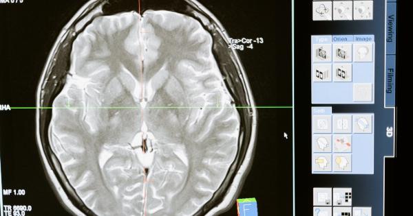
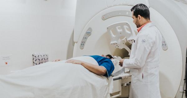
The first step in building a 3D-printed heart is acquiring accurate patient-specific anatomical data through medical imaging techniques such as computed tomography (CT) or magnetic resonance imaging (MRI).
These imaging technologies provide detailed information about the patient’s heart, including its size, shape, and structural abnormalities.
2. Creating a Digital Model
Once the imaging data is obtained, it is converted into a digital 3D model using specialized software. This digital model serves as the blueprint for the 3D printing process and guides the printing of each layer.
3. Material Selection
The choice of materials used for 3D printing a heart is critical to ensure the organ’s functionality, durability, and compatibility within the human body.
Biocompatible materials that possess properties similar to natural heart tissue, such as hydrogels or bioinks, are typically used in the printing process.
4. Printing the Heart
Using the digital model, the 3D printing process begins by depositing layers of the chosen material on a platform according to the predetermined design. The layers slowly build upon one another, gradually forming the structure of the heart.
The printing process requires precision and accuracy to ensure the creation of intricate features and a functional heart.
5. Post-Processing and Finishing
Once the heart is fully printed, it undergoes post-processing and finishing steps to refine its structure and enhance its functionality.
This may involve washing, curing, or annealing the printed structure to remove any residual materials or enhance its mechanical properties.
6. Integration of Blood Vessels
A crucial aspect of building a functional 3D-printed heart is the integration of blood vessels. Without proper blood supply, the heart cannot sustain its own function.
Researchers are exploring innovative techniques, such as embedding channels or using bioprinting methods, to create a network of blood vessels within the printed heart.
7. Cellularization and Tissue Maturation
To achieve a fully functional 3D-printed heart, researchers are working on methods to populate the printed structure with living cells.
Through a process called cellularization, stem cells or patient-specific cells are seeded onto the printed heart scaffold. The cells then proliferate and develop, maturing over time to form functional heart tissue.
8. Testing and Validation
Before a 3D-printed heart can be considered viable for transplantation, it undergoes rigorous testing and validation procedures.
Researchers conduct extensive anatomical, physiological, and functional assessments to ensure the organ’s performance meets the required standards.
9. Ethical Considerations
Building a heart with 3D printing technology poses ethical considerations. The potential for manufacturing human organs raises questions regarding organ availability, affordability, and equity in healthcare.
Researchers and policymakers must address these concerns to develop ethical guidelines and ensure equitable access to this revolutionary technology.
The Future of 3D-Printed Hearts
The development of 3D-printed hearts holds immense promise for the future of cardiac medicine. This technology has the potential to overcome the limitations posed by organ transplantation, reduce waiting times, and ultimately save countless lives.
Continued advancements in material science, cellular biology, and 3D printing technologies will further propel the field forward.
Conclusion
Building a heart with 3D printing technology represents a significant milestone in the quest to overcome the challenges of heart transplantation.
The ability to create fully-functional hearts using patient-specific anatomical data opens up a world of possibilities for customized treatment options and reduces the burden on organ donors. While challenges remain, the future of 3D-printed hearts looks promising, offering hope to those in need of life-saving interventions.


