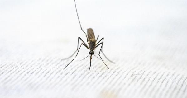
Malaria is a life-threatening disease caused by the Plasmodium parasite. It is transmitted to humans through the bites of infected female mosquitoes. Once inside the body, the parasites travel to the liver, mature, and then invade red blood cells.
This is when the symptoms of malaria start to manifest.
Understanding the importance of trapping parasites
Trapping and studying the malaria parasites in the blood is crucial for various reasons. Firstly, it helps in diagnosing the disease accurately.
By identifying the presence and the strain of the parasite, healthcare professionals can determine the appropriate treatment plan. Secondly, studying the parasites provides valuable information for vaccine development and drug resistance monitoring.
Lastly, trapping parasites is instrumental in understanding the complex life cycle of Plasmodium and its interactions with the human immune system.
Common techniques for trapping malaria parasites
Several techniques have been developed to trap malaria parasites in the blood. These methods range from traditional microscopic examination to more advanced molecular techniques.
1. Giemsa staining and microscopy
Giemsa staining is a widely used method for visualizing malaria parasites. Blood smears are prepared and stained with Giemsa solution, which selectively stains the parasite’s structures, making them visible under a microscope.
Microscopy allows for the identification and quantification of parasitized red blood cells. However, this technique has limitations in terms of sensitivity, especially at low parasitemia levels.
2. Rapid diagnostic tests (RDTs)
Rapid diagnostic tests are simple, easy-to-use tools for detecting malaria antigens in the blood.
These tests work on the principle of immunochromatography, where specific antibodies attached to test lines capture and bind to parasite or antigen-specific proteins. RDTs provide quick results and have high specificity but may have lower sensitivity compared to other methods.
3. Polymerase chain reaction (PCR)
Polymerase chain reaction is a molecular technique used to amplify and detect the genetic material (DNA or RNA) of the malaria parasite. PCR-based methods are highly sensitive and specific, capable of detecting even low levels of parasites in the blood.
They can also differentiate between different Plasmodium species and detect drug-resistant strains. However, PCR requires specialized equipment, skilled technicians, and is relatively expensive.
4. Loop-mediated isothermal amplification (LAMP)
LAMP is a newer molecular technique that amplifies DNA sequences under isothermal conditions. Unlike PCR, LAMP does not require sophisticated equipment and can be performed in simpler laboratory settings.
LAMP assays are highly sensitive, specific, and can detect low parasite densities. They also have the advantage of providing visual results, as LAMP-generated amplification products produce a color change.
5. Fluorescence microscopes
Fluorescence microscopes utilize fluorescent dyes that specifically bind to malaria parasites, making them easily distinguishable from normal red blood cells. This technique allows for rapid and accurate identification of parasitemia levels.
Fluorescence microscopy is particularly useful when studying drug resistance or monitoring treatment outcomes since it can assess the viability of parasites.
6. Microfluidic devices
Microfluidic devices are small, integrated systems that manipulate and analyze fluids at a microscopic scale. These devices can be designed to accommodate small volumes of blood and incorporate various techniques such as PCR or microscopy.
Microfluidic platforms offer faster processing times and reduced reagent requirements, making them suitable for field settings and resource-limited areas.
7. Ex vivo culture systems
Ex vivo culture systems involve growing malaria parasites obtained from patient blood samples in the laboratory.
This technique allows for the study of parasite biology, drug susceptibility testing, and testing the efficacy of potential antimalarial drugs. Culturing parasites ex vivo enables researchers to manipulate and control experimental conditions, providing valuable insights into the parasite’s behavior and response to different interventions.
8. Capture methods using magnetic beads
Capture methods involve using magnetic beads coated with specific antibodies that bind to malaria parasites. The beads are added to blood samples, and parasite-infected red blood cells are selectively captured and separated using magnetic fields.
This technique allows for enrichment and concentration of parasites, improving their detection and downstream analysis.
9. Image analysis and artificial intelligence
Advancements in image analysis and artificial intelligence have enabled the development of automated systems for malaria parasite detection.
These systems use sophisticated algorithms to analyze digital images of stained blood smears or other diagnostic tests. They can accurately identify and quantify parasites, eliminating the need for manual microscopy and reducing human error.
10. Flow cytometry
Flow cytometry is a technique that utilizes lasers and detectors to analyze the characteristics of individual cells as they pass through a flow cell.
It can identify and measure various parameters of malaria-infected red blood cells, such as parasitemia levels, cell size, and complexity. Flow cytometry provides rapid and high-throughput analysis, making it valuable for large-scale epidemiological studies.


