Stroke is a medical emergency that occurs when the blood supply to a part of the brain is interrupted or reduced, depriving brain tissue of oxygen and nutrients. It can lead to serious complications and even death if not treated promptly.
In the management of stroke, imaging tests play a crucial role in both diagnosis and treatment. These tests allow healthcare professionals to identify the type and location of the stroke, assess its severity, and guide appropriate interventions.
Angiography: Visualizing Blood Vessels
Angiography is a widely used imaging technique that allows doctors to visualize blood vessels in the brain. It can help in diagnosing the cause of a stroke, such as a blood clot or a weakened blood vessel.
During the procedure, a contrast agent is injected into the blood vessels, followed by a series of X-rays or computed tomography (CT) scans. By examining the images produced, healthcare professionals can identify any abnormalities, narrowings, or blockages in the blood vessels that may be contributing to the stroke.
CT Scan: Assessing Brain Damage
Computed tomography (CT) scan is another imaging test commonly used in stroke management. It involves taking a series of X-ray images of the brain from different angles, which are then processed by a computer to create detailed cross-sectional images.
CT scans can quickly assess the extent of brain damage caused by a stroke, determine the type of stroke (ischemic or hemorrhagic), and rule out other conditions with similar symptoms. Additionally, CT angiography can provide detailed images of the blood vessels to aid in the diagnosis and planning of potential interventions.
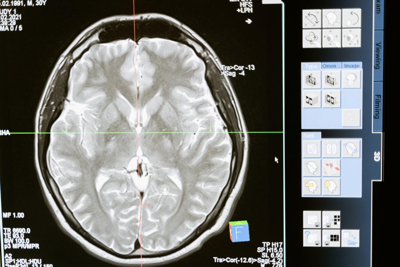
MRI: Detailed Visualization
Magnetic resonance imaging (MRI) is a non-invasive imaging test that uses powerful magnets and radio waves to produce detailed images of the brain.
It provides more detailed visualization of the brain structures and is particularly useful in detecting small strokes or brain lesions that may not be clearly visible on a CT scan. MRI can help determine the age of a stroke, identify the affected brain regions, and assess the viability of brain tissue. It is also used to monitor the progress of stroke patients over time and evaluate the effectiveness of treatment strategies.
Doppler Ultrasound: Assessing Blood Flow
Doppler ultrasound is a safe and painless imaging technique that uses sound waves to assess blood flow through the blood vessels.
It can be used to evaluate the blood flow in the arteries supplying the brain, check for any blockages or narrowing, and assess the degree of blood vessel damage. Doppler ultrasound can also be used to evaluate the blood flow within the brain itself, providing valuable information about the circulation in the affected areas.
This information is crucial in determining the appropriate treatment options for stroke patients.
Echocardiogram: Assessing Heart Function
Echocardiography, or echo, is a non-invasive test that uses sound waves to create detailed images of the heart. It can help evaluate the heart’s structure and function, identify any abnormalities, and assess the risk of stroke.
Related Article
Preventive Measures for Stroke: Imaging Techniques

In some cases, stroke may be caused by a blood clot that originates in the heart and travels to the brain. An echocardiogram can help identify such clots, as well as detect any other heart conditions that may contribute to the development of a stroke. This information guides the selection of appropriate treatment options and preventive measures.
Penumbra Imaging: Assessing Perfusion
Penumbra imaging is a relatively newer technique that assesses brain perfusion, or the blood supply to the brain tissue. It provides vital information about which areas of the brain are salvageable and which are irreversibly damaged.
By identifying the penumbra, or the area of brain tissue with reduced blood flow but still potentially salvageable, healthcare professionals can make informed decisions about reperfusion therapies, such as thrombolytic treatment or mechanical clot retrieval. Penumbra imaging plays a crucial role in determining the eligibility of stroke patients for time-sensitive interventions.
Stroke Management and Treatment Decisions
Imaging tests not only aid in the diagnosis of stroke but also guide treatment decisions. The information obtained from these tests helps healthcare professionals determine the most appropriate course of action for each individual patient.
For example, if the imaging tests reveal a blood clot causing an ischemic stroke, thrombolytic therapy or mechanical clot retrieval may be considered as potential interventions. On the other hand, if a hemorrhagic stroke is identified, immediate surgical interventions or medical management may be necessary to control bleeding and reduce further damage.
Furthermore, imaging tests are used to monitor patients during their recovery process.
They allow healthcare professionals to assess the effectiveness of treatment strategies, detect any new complications, and make necessary adjustments to the management plan. Follow-up imaging also helps in determining the long-term prognosis and identifying any secondary causes of stroke that need to be addressed, such as underlying vascular abnormalities or cardiac conditions.
Advancements in Imaging Technology
Advancements in imaging technology have significantly improved the accuracy and speed of stroke diagnosis and treatment.
Newer techniques, such as diffusion-weighted imaging (DWI) and perfusion-weighted imaging (PWI), provide valuable insights into the extent of brain damage, the affected tissue’s perfusion status, and the potential for recovery. These advanced imaging techniques help choose appropriate treatment options while minimizing the risk of complications.
Conclusion
Imaging tests play a critical role in controlling stroke by enabling healthcare professionals to accurately diagnose and assess the severity of the condition.
Various imaging techniques, including angiography, CT scan, MRI, Doppler ultrasound, echocardiogram, and penumbra imaging, offer valuable information about the cause, location, and extent of brain damage. These tests guide treatment decisions, help monitor the efficacy of interventions, and contribute to long-term management strategies. Advancements in imaging technology continue to revolutionize stroke care, enabling better outcomes for patients.


