Eye cancer, also known as ocular cancer, is a rare but potentially serious condition that occurs when abnormal cells grow uncontrollably in the tissues of the eye.
Early detection of eye cancer is crucial for effective treatment and improved patient outcomes. Medical imaging techniques such as photography, ultrasonography, and optical coherence tomography (OCT) play a significant role in the diagnosis and monitoring of this condition.
In this article, we will explore the educational images used for eye cancer detection.
1. Fundus Photography
Fundus photography, also known as retinal photography, is a non-invasive imaging technique that captures detailed images of the back of the eye, including the retina, optic disc, blood vessels, and macula.
These images can provide valuable information about any abnormalities or changes in the eye that may indicate the presence of cancerous growths or tumors.
2. Fluorescein Angiography
Fluorescein angiography is a diagnostic test that involves injecting a fluorescent dye into the bloodstream, which highlights the blood vessels in the retina.
This imaging technique helps ophthalmologists detect abnormalities in the blood vessels, identify tumor growth, and evaluate the extent of the disease in eye cancer patients.
3. Optical Coherence Tomography (OCT)
Optical coherence tomography (OCT) is a non-invasive imaging technique that uses light waves to create detailed cross-sectional images of the structures within the eye.
By providing high-resolution images of tissues at a cellular level, OCT can help ophthalmologists identify cancerous tumors and assess their size, location, and depth.
4. Ultrasound Imaging
Ultrasound imaging, also known as sonography, is a technique that uses sound waves to create images of the inside of the body.
In the context of eye cancer detection, ultrasound can be used to assess the thickness of the tumor, its location within the eye, and any changes in the surrounding tissues. This imaging modality is especially useful when there are opacities that hinder a clear view of the eye’s structures.
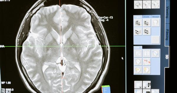
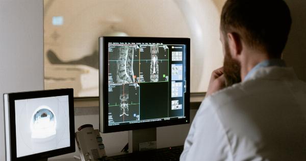
5. Magnetic Resonance Imaging (MRI)
Magnetic resonance imaging (MRI) is a powerful imaging technique that uses a magnetic field and radio waves to create detailed images of the body’s internal structures.
In the case of eye cancer, MRI can provide information about the size, extent, and spread of the tumor, as well as any involvement of adjacent structures such as the optic nerve or brain.
6. Computed Tomography (CT) Scan
Computed tomography (CT) scans use X-rays and computer processing to create detailed cross-sectional images of the body. This imaging technique can be beneficial in determining the size, location, and spread of eye cancer tumors.
CT scans can also help identify any metastases or spread of cancer to other areas of the body.
7. Infrared Retinal Imaging
Infrared retinal imaging involves capturing images of the eye using infrared light. This imaging technique can help identify changes in blood vessels, detect tumors, and monitor the response to treatment.
Infrared imaging is particularly useful for detecting specific types of eye cancer, such as retinoblastoma.
8. Autofluorescence Imaging
Autofluorescence imaging measures the natural fluorescence emitted by molecules in the eye.
This imaging technique can help identify abnormal changes in the eye’s tissues, including the presence of tumors or abnormal cell growth associated with eye cancer. Autofluorescence imaging is often used to detect and monitor retinal pigment epithelial tumors.
9. Confocal Scanning Laser Ophthalmoscopy (CSLO)
Confocal scanning laser ophthalmoscopy (CSLO) is an advanced imaging technique that uses laser light to obtain high-resolution images of the retina, optic nerve, and surrounding structures.
These images can be used to detect and monitor eye tumors, as well as assess the effectiveness of treatment interventions.
10. Corneal Topography
Corneal topography is a specialized imaging technique that maps the curvature and shape of the cornea, the clear outer layer of the eye.
This imaging modality can help identify any irregularities or changes in the cornea that may be associated with certain types of eye cancer, such as ocular surface squamous neoplasia.


