Radiographs, also known as X-rays, have become an integral part of medical diagnostics. These imaging techniques use ionizing radiation to capture images of internal structures in the human body.
However, concerns have been raised about the potential link between radiographs and an increased risk of cancer. In this article, we will examine the available research on this topic to determine if there is indeed a connection between radiographs and cancer risk.
Understanding Radiographs
Radiographs are a valuable tool in medical imaging as they can help diagnose various conditions and monitor treatment progress. They work by passing a controlled amount of radiation through the body, which is then captured on a special film or detector.
This creates an image that reveals the internal structures, such as bones and organs.
Radiographs have been used for decades, and their benefits in diagnosing and treating diseases are well-known. However, the potential harm caused by exposure to ionizing radiation has raised concerns, especially when it comes to the risk of cancer.
The Link between Ionizing Radiation and Cancer
Ionizing radiation, including that used in radiographs, has the potential to damage DNA within cells. This damage can lead to mutations and disruptions in the cell’s normal functioning, potentially increasing the risk of cancer development.
While the harmful effects of high doses of radiation are well-established, the risk associated with the low doses typically used in radiographs is much less clear.
Extensive research has been carried out to understand the potential link between radiographs and cancer risk.
Evidence from Epidemiological Studies
Epidemiological studies play a crucial role in investigating the link between radiographs and cancer. These studies analyze large populations over extended periods to assess the association between exposure to radiation and the development of cancer.
Several such studies have been conducted, focusing on different populations and types of cancer.
Most of these studies have found a slightly increased risk of certain cancers, such as leukemia and thyroid cancer, in individuals who have been exposed to radiation from radiographs. However, it is important to note that the absolute risks are generally very low.
One notable study is the Life Span Study of Japanese Atomic Bomb Survivors, which followed a population exposed to high doses of radiation from atomic bombs.
The study found an increased risk of various cancers, including lung, breast, and stomach cancers, in those exposed to radiation. However, it is important to note that the doses received by the survivors were significantly higher than those associated with radiographs.
Factors Influencing Cancer Risk
It is important to consider various factors that can influence the potential cancer risk associated with radiographs. One such factor is the cumulative dose of radiation received over time.
The risk of cancer is thought to increase with higher cumulative doses, so individuals who frequently undergo radiographs may be at a slightly higher risk than those who have fewer procedures.
Another factor is the age at exposure. Children and young adults are generally considered more susceptible to the effects of radiation due to their rapidly dividing cells.
Therefore, the potential risk associated with radiographs may be higher for these age groups compared to older individuals.
Radiation Safety Measures
Over the years, significant efforts have been made to improve radiation safety in medical settings.
Guidelines and protocols have been developed to ensure that radiographs are only carried out when necessary and using the lowest possible dose of radiation.
Technological advancements have also contributed to reducing radiation doses. Modern X-ray machines are designed to be more efficient and precise, limiting unnecessary exposure.
Moreover, radiology technicians are trained to position patients correctly to minimize radiation exposure to non-targeted areas.
Other Imaging Alternatives
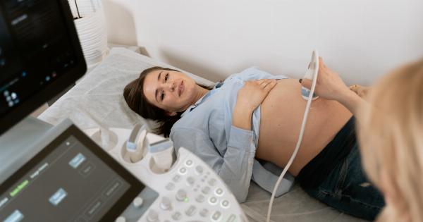
While radiographs are valuable, other imaging techniques, such as ultrasound and magnetic resonance imaging (MRI), can often provide similar diagnostic information without the use of ionizing radiation.
Ultrasound uses sound waves to generate images, making it a safe option for evaluating various conditions. MRI, on the other hand, relies on magnetic fields and radio waves to create detailed images of the body’s structures.
These alternative imaging methods may be preferred in situations where the risk associated with radiation needs to be avoided or minimized.
Conclusion
While radiographs have undeniably revolutionized medical diagnostics, concerns about their potential link to cancer risk have arisen.
The available evidence suggests that exposure to radiographs may slightly increase the risk of certain cancers, though absolute risks remain low. Factors such as cumulative dose, age at exposure, and adherence to radiation safety measures play a role in determining the potential risk.
It is important for medical professionals to weigh the benefits of radiographs against the potential risks and consider alternative imaging methods, especially in situations where the use of ionizing radiation can be avoided.
Further research and ongoing efforts to improve radiation safety measures are crucial to minimize any potential harm associated with radiographs.


