With the advancements in medical imaging technology, researchers and healthcare professionals have gained a powerful tool to better understand and diagnose various diseases.
Putpore images, in particular, have become increasingly popular in disease exploration due to their ability to provide detailed visual representations of tissues and organs. Through the analysis of these images, valuable insights can be gained, leading to improved disease detection, treatment, and prevention strategies.
The Power of Putpore Imaging
Putpore imaging is a non-invasive technique that allows medical professionals to capture images of internal body structures.
It utilizes special equipment, such as X-rays, ultrasounds, magnetic resonance imaging (MRI), computed tomography (CT) scans, and positron emission tomography (PET) scans, to generate detailed visual representations. These images offer valuable insights into the human body, enabling researchers to explore disease links and develop effective interventions.
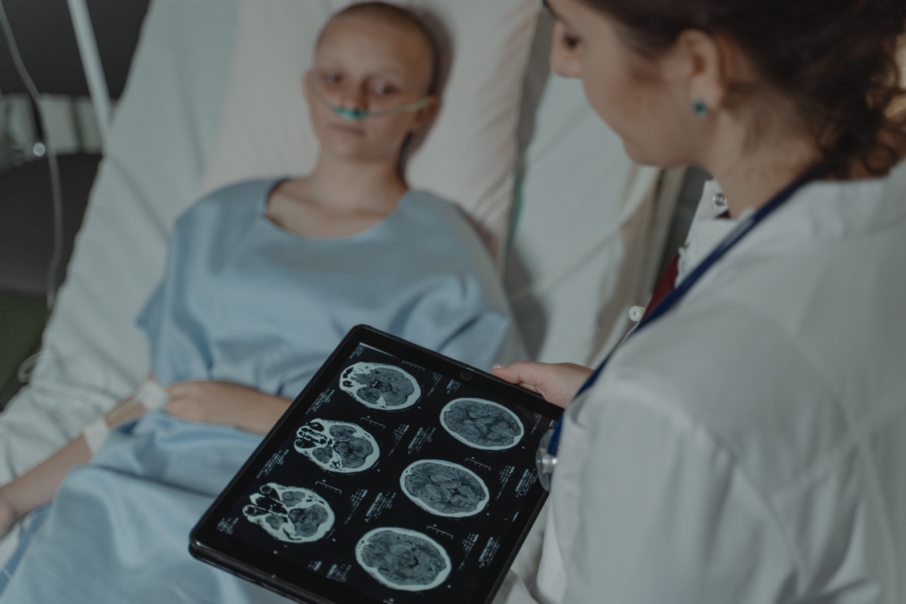
Diagnosing Diseases with Putpore Images
Putpore images are widely utilized in diagnosing various diseases.
The information obtained from these images provides doctors with a clearer understanding of the patient’s condition, assisting in the identification of abnormalities or potential disease risks. For example, chest X-rays help diagnose respiratory conditions such as pneumonia, lung cancer, or tuberculosis, while MRI scans are useful in detecting brain tumors, multiple sclerosis, or spinal cord injuries.
In addition to identifying specific diseases, putpore images also aid in disease staging, which helps determine the severity and progression of a condition.
This information is crucial for developing appropriate treatment plans and assessing the effectiveness of interventions.
Exploration of Disease Links
Putpore images offer a unique opportunity for researchers to explore disease links, providing valuable information for disease prevention and management.
By analyzing a large collection of images, patterns and similarities between different disease types can be identified. These findings enable medical professionals to better understand the underlying causes and mechanisms of various diseases, leading to improved strategies for prevention, early detection, and treatment.
For example, using sophisticated image analysis algorithms, researchers have discovered connections between cardiovascular diseases and certain artery patterns visible in angiograms.
This insight has allowed for advancements in predicting an individual’s risk for cardiovascular issues and developing personalized prevention strategies.
Advancements in Putpore Imaging Technology
The field of putpore imaging is constantly evolving, with new technologies and techniques being developed to improve disease exploration. One notable advancement is the incorporation of artificial intelligence (AI) algorithms in image analysis.
Related Article
Putpore: Diseases Linked with Pictorial Representations

AI algorithms can analyze large volumes of putpore images, identifying subtle patterns and abnormalities that may not be readily apparent to the human eye.
These AI-assisted analyses provide a more efficient and accurate diagnosis, enabling doctors to make informed decisions regarding disease management.
AI algorithms have demonstrated promising results in various fields, including cancer detection, cardiovascular disease assessment, and brain anomaly identification.
Challenges and Limitations
While putpore imaging is a powerful tool in disease exploration, there are challenges and limitations that need to be addressed.
One of the primary limitations is the high cost associated with imaging equipment and the expertise required to operate and interpret the results. This restricts widespread access to advanced putpore imaging technologies, particularly in resource-limited settings.
Another challenge is the need for a standardized approach to image analysis and interpretation. This ensures consistency and reliability across different healthcare institutions and facilitates the sharing of data and findings.
Efforts are underway to develop standardized protocols and guidelines for putpore imaging analysis, aiming to enhance collaboration and accelerate discoveries.
Future Possibilities
The future of disease exploration through putpore images holds great promise. As technology continues to advance, we can expect further improvements in imaging resolution, accuracy, and accessibility.
This will open up new possibilities for disease research, potentially leading to breakthroughs in early detection, targeted therapies, and personalized medicine.
Furthermore, the integration of putpore imaging with other medical data sources, such as genomic information or electronic health records, can provide a comprehensive understanding of disease progression, individual risk factors, and treatment outcomes. This interdisciplinary approach has the potential to revolutionize the field of medicine, leading to more precise and effective disease management strategies.
In Conclusion
Putpore imaging offers a window into the intricate workings of the human body, allowing researchers and healthcare professionals to explore disease links and develop improved strategies for prevention, diagnosis, and treatment.
The continuous advancements in putpore imaging technology, coupled with the integration of artificial intelligence, provide exciting possibilities for the future of disease exploration. By harnessing the power of putpore images, we can unlock vital insights that will ultimately improve patient outcomes and enhance overall healthcare practices.


