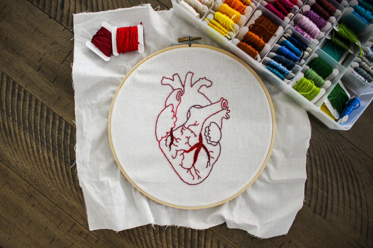
The heart, this vital organ that pumps blood throughout the body, is often referred to as the center of our being. It works tirelessly day in and day out, ensuring the delivery of oxygen and nutrients to every cell.
Understanding the anatomy of the heart is crucial in comprehending its complex functioning. In this article, we will take a journey into the intricate structure of the heart and explore its various components.
The Size and Location of the Heart
The heart is approximately the size of a closed fist and is located within the thoracic cavity, in a slightly offset position towards the left side. It rests between the lungs, just behind the breastbone (sternum), and is protected by the rib cage.
The Pericardium
The heart is surrounded by a protective double-layered sac called the pericardium.
The tough outer layer known as the fibrous pericardium provides stability and prevents overfilling of the heart, while the inner layer called the serous pericardium helps reduce friction as the heart beats.
Chambers of the Heart
The heart is divided into four chambers: two atria and two ventricles. The atria are the upper chambers, and their main function is to receive blood from the body (right atrium) and the lungs (left atrium).
The ventricles, on the other hand, are the lower chambers responsible for pumping blood out of the heart into the circulatory system.
The Atria
The atria are smaller and thinner-walled compared to the ventricles. They primarily act as holding chambers, allowing blood to flow into them before it is propelled into the ventricles.
The right atrium receives deoxygenated blood from the body via two main veins: the superior vena cava and the inferior vena cava. The left atrium receives oxygenated blood from the lungs through four pulmonary veins.
The Ventricles
The ventricles are the powerhouse of the heart and are responsible for pumping blood to the rest of the body.
The right ventricle pumps deoxygenated blood to the lungs for oxygenation, while the left ventricle pumps oxygenated blood to the various organs and tissues. The walls of the ventricles are thicker and more muscular than those of the atria to support their robust pumping action.
Valves of the Heart
The heart contains four valves that ensure smooth and uni-directional blood flow through its chambers.
The tricuspid valve separates the right atrium from the right ventricle, while the mitral valve (or bicuspid valve) separates the left atrium from the left ventricle. Both these valves open to allow blood flow from the atria to the ventricles and close to prevent backflow.
The pulmonary valve guards the opening between the right ventricle and the pulmonary artery. When the ventricle contracts, the pulmonary valve opens to allow blood to be pumped into the pulmonary artery for oxygenation.
Finally, the aortic valve is situated between the left ventricle and the aorta, the large artery that carries oxygenated blood to the rest of the body. The aortic valve opens when the left ventricle contracts, enabling blood to flow into the aorta.
The Cardiac Cycle
The cardiac cycle refers to the sequence of events that occur during one complete heartbeat. It consists of two phases: systole and diastole. During systole, the ventricles contract, pumping blood out of the heart into the arteries.
On the other hand, diastole is the relaxation phase when the ventricles refill with blood. This continuous cycle ensures the constant circulation of blood throughout the body.
Coronary Arteries
The heart, like any other muscle, requires a constant supply of oxygen and nutrients to function properly. This is made possible by the coronary arteries, which branch off from the root of the aorta and encircle the heart.
The left coronary artery gives rise to the left anterior descending artery (LAD) and the left circumflex artery, supplying blood to different areas of the heart. The right coronary artery supplies blood to the right atrium and ventricle, as well as to the back of the heart.
Electrical Conduction System of the Heart
Contrary to popular belief, the heart does not rely on external stimulation to contract. It possesses its own built-in electrical conduction system, ensuring a coordinated and rhythmic heartbeat.
The sinoatrial (SA) node, often called the heart’s natural pacemaker, generates electrical impulses that initiate each heartbeat. These impulses then travel through the atria and reach the atrioventricular (AV) node, located at the junction between the atria and ventricles.
The AV node acts as a bridge, delaying the impulses slightly before transmitting them to the ventricles via specialized fibers known as the bundle of His and Purkinje fibers.
Conclusion
The human heart, with its remarkable anatomy and physiology, is undoubtedly a marvel of creation.
From its relentless pumping action to its intricate network of vessels and valves, every component works together to ensure the continuous circulation of blood throughout the body. Understanding the anatomy of the heart not only deepens our appreciation for this vital organ but also enhances our awareness of the importance of maintaining heart health.
