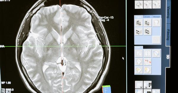
Meningiomas are slow-growing tumors that develop in the meninges, which are the protective layers of tissue that surround the brain and spinal cord.
These tumors are usually noncancerous (benign), but in some cases, they can become cancerous (malignant). Diagnosing meningiomas is crucial for determining the appropriate treatment plan and ensuring the best possible outcome for the patient. This article will explore the various methods used to diagnose meningiomas.
1. Medical history and physical examination
The diagnostic process for meningiomas typically begins with a detailed medical history and physical examination. The doctor will inquire about the patient’s symptoms, family medical history, and any previous medical conditions or surgeries.
During the physical examination, the doctor will assess the patient’s cognitive function, sensory abilities, reflexes, and coordination.
2. Neurological examination
A neurological examination is a crucial step in diagnosing meningiomas. The doctor will evaluate the patient’s nervous system function, focusing on any abnormalities that may indicate the presence of a brain tumor.
This examination may include tests such as checking reflexes, assessing muscle strength and coordination, and assessing sensory function.
3. Imaging tests
Imaging tests are essential for diagnosing meningiomas and determining their size, location, and characteristics. The most commonly used imaging techniques include:.
a. Magnetic Resonance Imaging (MRI)
MRI scans use powerful magnets and radio waves to create detailed images of the brain and spinal cord. This noninvasive imaging test provides high-resolution images, allowing doctors to visualize meningiomas and assess their characteristics.
b. Computed Tomography (CT) scan
CT scans use X-rays and computer technology to produce detailed cross-sectional images of the brain.
This imaging technique can help identify meningiomas, detect any associated bleeding or bone abnormalities, and assess the tumor’s size and location.
c. Angiogram
An angiogram involves injecting a contrast dye into blood vessels to make them visible on X-ray images. Angiograms can help identify abnormal blood vessels and assess the blood supply to meningiomas. This information is crucial for surgical planning.
d. Positron Emission Tomography (PET) scan
A PET scan uses a small amount of radioactive material to detect increased cell activity in the body. This imaging technique can help differentiate between benign and malignant meningiomas, as malignant tumors often show increased cell metabolism.
4. Biopsy
A biopsy involves removing a small sample of tissue for laboratory analysis. While a definitive diagnosis of meningioma can often be made based on imaging tests, a biopsy may be necessary to confirm the diagnosis or assess the tumor’s grade.
There are two types of biopsies commonly used for meningiomas:.
a. Needle biopsy
A needle biopsy involves inserting a thin needle through the skin and into the tumor to obtain a tissue sample. This procedure is usually guided by imaging techniques such as MRI or CT scans.
Needle biopsies are less invasive than surgical biopsies and can be performed on tumors located in challenging areas.
b. Surgical biopsy
In some cases, a surgical biopsy may be necessary. This involves removing a larger sample of the tumor during a surgical procedure.
Surgical biopsies are typically performed when the tumor is accessible or when there is uncertainty about its nature based on imaging tests.
5. Genetic testing
Genetic testing may be recommended for individuals with meningiomas, particularly those with multiple or recurring tumors.
These tests can help identify specific gene mutations associated with certain types of meningiomas, allowing for more targeted treatment approaches.
6. Spinal tap (Lumbar puncture)
In rare cases, a spinal tap may be performed to analyze the cerebrospinal fluid for signs of meningioma. This procedure involves inserting a needle into the lower back to collect a small sample of fluid surrounding the spinal cord.


