Varicose veins are a common condition that affects many people, especially women. These dilated, twisted veins can cause discomfort, pain, swelling, and even skin changes. While some varicose veins are purely a cosmetic issue, others require treatment.
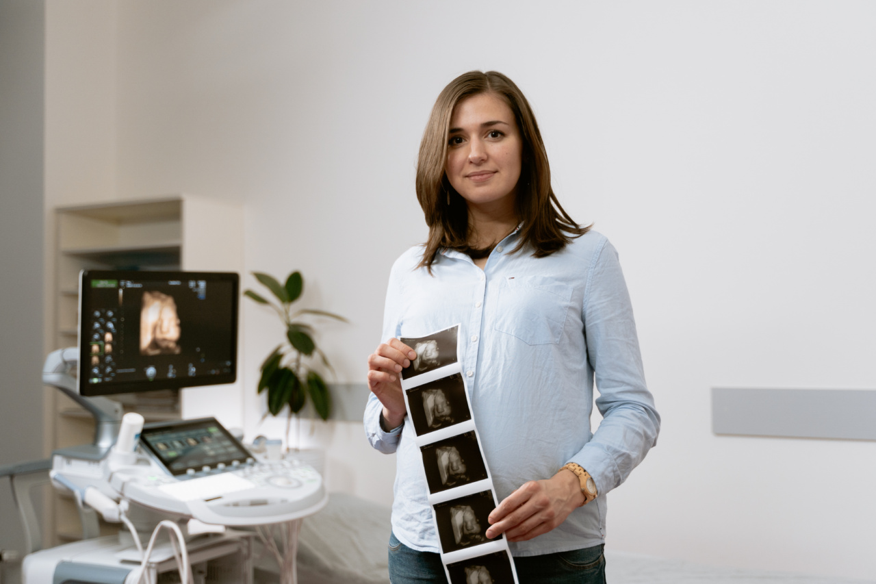
Proper diagnosis is crucial to determine the extent of the problem and its underlying causes. One way to diagnose varicose veins is through ultrasound imaging, a non-invasive technique that uses sound waves to create images of the veins and blood flow.
What Are Varicose Veins and Their Symptoms?
Varicose veins occur when the valves in the veins that regulate blood flow from the legs to the heart are damaged or weakened, causing blood to pool and enlarge the veins. The most common symptoms of varicose veins include:.
- Bulging veins that are blue, purple, or red in color
- Twisted or rope-like veins
- Pain or aching in the legs, especially after standing or sitting for long periods
- Swelling in the legs, ankles, or feet
- Heaviness or fatigue in the legs
- Burning or itching sensations
- Ulcers or open sores in severe cases
What Is Ultrasound Imaging?
Ultrasound imaging, also known as sonography, uses high-frequency sound waves to create images of the body’s internal structures, including the organs, blood vessels, and tissues.
This technique is safe, painless, and non-invasive, making it ideal for diagnosing various conditions without the need for surgery or invasive procedures. Ultrasound imaging is widely used in obstetrics to monitor fetal development, as well as in cardiology, gastroenterology, and other medical specialties.
How Does Ultrasound Imaging Diagnose Varicose Veins?
To diagnose varicose veins, an ultrasound technician or radiologist uses a handheld device called a transducer to emit sound waves into the leg and capture the echoes that bounce back.
These echoes are then converted into images on a computer screen, which the doctor can interpret. Ultrasound imaging can show the size, location, and shape of the veins, as well as the direction and speed of the blood flow.
This information helps the doctor determine if the valves in the veins are functioning properly, or if there are any blockages or blood clots that require further evaluation.
Related Article
Understanding varicose veins through ultrasound imaging

What Types of Ultrasound Imaging Are Used for Varicose Veins?
There are two main types of ultrasound imaging used for varicose veins:.
Doppler Ultrasound
Doppler ultrasound is a type of ultrasound that shows the direction and speed of blood flow in the veins.
This technique uses a special software that translates the sound waves into a color-coded display on the monitor, with red indicating blood flow towards the transducer and blue representing flow away from the transducer. Doppler ultrasound is particularly useful in evaluating the blood flow in the deep veins, as well as the superficial veins. This technique can also detect blood clots or other obstructions that might be causing the varicose veins.
Duplex Ultrasound
Duplex ultrasound is a combination of regular ultrasound and Doppler ultrasound. This technique can show both the structure of the veins and the blood flow within them.
Duplex ultrasound can reveal varicose veins that are not visible on the surface, as well as assess the severity of the problem. The doctor can also use this technique to plan the appropriate treatment, whether it’s conservative measures like wearing compression stockings or more invasive options like endovenous laser treatment or phlebectomy.
What Are the Benefits of Ultrasound Imaging for Varicose Veins?
Ultrasound imaging has several advantages when it comes to diagnosing and treating varicose veins:.
- It’s non-invasive, with no needles or incisions required
- It’s painless and safe, with no exposure to radiation
- It’s accurate and precise, providing detailed images of the veins and blood flow
- It allows for early detection and intervention, which can prevent complications
- It guides the treatment plan and helps monitor the progress of the treatment
- It’s cost-effective and widely available
Conclusion
Ultrasound imaging is a valuable tool for diagnosing varicose veins and assessing their severity. This non-invasive technique enables doctors and technicians to visualize the veins, measure blood flow, and identify any underlying conditions.
With proper diagnosis and treatment, patients with varicose veins can improve their symptoms, prevent complications, and enjoy better quality of life. If you’re experiencing any symptoms of varicose veins, talk to your doctor about the benefits of ultrasound imaging and other diagnostic tests.


