Pyelonephritis is a type of kidney infection that typically occurs as a result of a urinary tract infection (UTI). It is important to promptly diagnose and treat pyelonephritis to prevent further complications.
In addition to clinical evaluation and laboratory tests, medical imaging plays a crucial role in identifying the presence of pyelonephritis and assessing the extent of kidney involvement. This article aims to provide an overview of the key signs to look for in imaging studies when diagnosing pyelonephritis.
1. Clinical Presentation:
Before delving into the imaging findings, it is essential to recognize the symptoms and clinical features of pyelonephritis. Common signs and symptoms include:.
- Fever and chills
- Flank pain or tenderness
- Abdominal pain
- Frequent urination
- Urinary urgency
- Blood in urine (hematuria)
- Cloudy or foul-smelling urine
2. CT Scan:
Computed Tomography (CT) scan is one of the most commonly used imaging techniques to visualize the kidneys and identify signs of pyelonephritis.
CT scans provide detailed cross-sectional images of the abdomen and pelvis, which are helpful in evaluating the kidneys and detecting any abnormal findings.
Key findings indicative of pyelonephritis on a CT scan include:.
- Enlarged kidneys
- Perinephric stranding (inflammation and edema around the kidneys)
- Presence of abscesses or fluid collections in or around the kidneys
- Thickening of the renal pelvis or walls of the ureters
- Hydronephrosis (enlargement of the kidney due to obstruction)
3. Ultrasound:
Ultrasound imaging is another valuable tool for assessing and diagnosing pyelonephritis. It is non-invasive and does not involve exposure to radiation, making it a safer option, especially for pregnant women and younger patients.
Some characteristic ultrasound findings suggestive of pyelonephritis include:.
- Increased echogenicity (brightness) of the renal parenchyma
- Presence of hypoechoic areas (dark regions) within the kidneys
- Enlarged kidneys
- Dilated or thickened ureters
- Loss of corticomedullary differentiation (normal distinction between the outer and inner regions of the kidney)
- Pelvicalyceal dilatation (enlarged renal collecting system)
4. X-ray:
X-rays may be used to evaluate pyelonephritis, although they are less sensitive and specific compared to CT scans or ultrasounds. However, they can help identify certain associated complications such as kidney stones or structural abnormalities.
Key X-ray findings in pyelonephritis may include:.
- Calyceal or ureteric calcifications (stones)
- Enlarged or distorted kidneys
- Abscesses or gas within the kidney
5. MRCP (Magnetic Resonance Cholangiopancreatography):
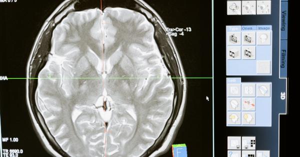
In some cases, magnetic resonance imaging (MRI) with MRCP may be utilized to assess the kidneys and urinary tract in detail.
MRCP helps visualize the renal system’s anatomy, including the collecting ducts and ureters, and may provide additional information about any obstructive or structural abnormalities contributing to pyelonephritis.
Common MRCP findings in pyelonephritis include:.
- Enlarged kidneys due to inflammation
- Presence of abscesses or fluid collections
- Evidence of obstructive uropathy
Conclusion:
Imaging techniques such as CT scans, ultrasound, X-rays, and MRCP play a crucial role in diagnosing pyelonephritis.
It is important to look for specific signs and findings in these imaging studies to confirm the presence of kidney infection and assess the severity of the disease. Early detection and appropriate treatment can help prevent complications and ensure a favorable outcome for patients with pyelonephritis.


