Strokes are medical emergencies that occur when the blood supply to the brain is interrupted or reduced. Rapid diagnosis and treatment are instrumental in preventing permanent brain damage, disability and even death.
Brain imaging techniques play a crucial role in the diagnosis, treatment and prevention of strokes. This article will discuss the importance of imaging in stroke prevention and the different types of imaging that are used.
Why Imaging is Important in Stroke Prevention
Imaging is important in stroke prevention because it can help identify risk factors, diagnose a stroke, help decide the best treatment plan and monitor the success of treatment.
It can also provide information on the size, location and severity of the stroke. Accurate diagnosis and treatment is essential in preventing another stroke from happening in the future.
Types of Imaging Used in Stroke Prevention
Here are some of the different imaging techniques used in stroke prevention:.
CT Scan
CT scans use X-rays to produce detailed images of the brain. They can detect changes such as bleeding or brain swelling that may be caused by a stroke.
CT scans are usually the first imaging technique used in a stroke emergency because they are fast and can quickly identify bleeding or other life-threatening conditions.
MRI
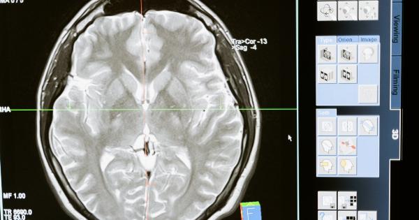
MRI uses magnetic fields and radio waves to produce detailed pictures of the brain. It is more sensitive than a CT scan and can detect changes in brain tissue caused by a stroke earlier.
MRI is also useful in identifying small areas of damage which may be missed by a CT scan.
Cerebral Angiography
Cerebral angiography is an imaging technique that uses X-rays and a special dye injected into the blood vessels to examine the arteries that supply blood to the brain.
It can identify narrow or blocked blood vessels in the brain and detect aneurysms or other vascular abnormalities. Cerebral angiography is not usually used as the first test, but can be helpful in planning treatment and preventing future strokes.
Ultrasound
Ultrasound uses sound waves to produce images of the brain. It is a noninvasive technique that can detect changes in blood flow and identify narrowing or blockage in the arteries that supply blood to the brain.
Ultrasound can also detect blood clots that may have caused a stroke.
Transcranial Doppler
Transcranial Doppler is a type of ultrasound that uses sound waves to measure the speed and direction of blood flow in the arteries in the brain.
It can detect changes in blood flow caused by narrowing or blockages in the arteries and can help identify patients at risk of a stroke.
Conclusion
Imaging plays a crucial role in stroke prevention by assisting in the diagnosis, treatment and monitoring of strokes. It helps identify risk factors, diagnose a stroke and the underlying cause and help make treatment decisions.
Patients who have had a stroke or are at risk of a stroke should discuss imaging and treatment options with their doctors.


