Gynecological cancers affect a significant number of women worldwide, with early detection being crucial for successful treatment outcomes.
Imaging techniques play a vital role in the basic prevention of gynecological cancer by aiding in early detection, accurate diagnosis, and monitoring of the disease. This article explores various imaging techniques used in the prevention of gynecological cancer.
Pap Smear Test
The Pap smear test, or Pap test, is a widely used screening tool for the detection of cervical cancer. It involves collecting cells from the cervix and examining them under a microscope to identify any abnormal changes.
By detecting precancerous or cancerous cells early on, the Pap smear test enables timely interventions and reduces the risk of developing advanced-stage cervical cancer.
Transvaginal Ultrasound
Transvaginal ultrasound is an imaging technique that uses high-frequency sound waves to create detailed images of the female reproductive organs.
By inserting a small probe into the vagina, images of the uterus, cervix, ovaries, and surrounding structures can be obtained. This technique helps in diagnosing ovarian and uterine cancers, as well as detecting abnormalities such as cysts or fibroids.
Mammography
Mammography is a specific type of breast imaging technique used for the early detection of breast cancer. It involves compressing the breast tissue and taking X-ray images to identify any abnormal masses or microcalcifications.
Regular mammograms are recommended for women over the age of 40, as they greatly contribute to the prevention and early diagnosis of breast cancer.
Colposcopy
Colposcopy is a procedure that allows a closer examination of the cervix, vagina, and vulva using a specialized magnifying instrument called a colposcope. It is often performed after an abnormal Pap smear or to further investigate suspicious areas.
Colposcopy aids in the detection of precancerous or cancerous changes in the cervix, guiding additional diagnostic and treatment procedures.
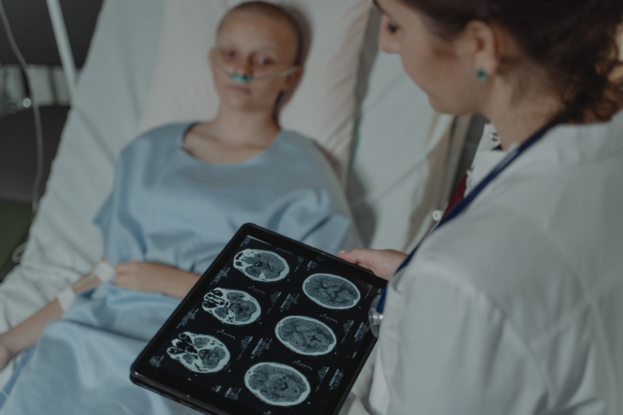
Computed Tomography (CT) Scan
Computed Tomography (CT) scan is a non-invasive imaging technique that uses X-rays to create detailed cross-sectional images of the body.
Related Article
Preventive Measures for Gynecological Cancer: Image-based Examination

It is commonly used to detect and stage gynecological cancers, as it provides a comprehensive view of the pelvis and surrounding structures. CT scan helps in determining the extent of tumor spread, guiding treatment planning decisions.
Magnetic Resonance Imaging (MRI)
Magnetic Resonance Imaging (MRI) is a powerful imaging technique that uses a magnetic field and radio waves to generate detailed images of the body.
In gynecological cancer, MRI helps in precise tumor localization, assessing tumor characteristics, and evaluating lymph node involvement. It is particularly valuable in detecting and monitoring ovarian and uterine cancers.
Positron Emission Tomography (PET) Scan
Positron Emission Tomography (PET) scan is a specialized imaging technique that provides functional information about tissues and organs.
It involves the injection of a small amount of radioactive tracer, which is taken up by rapidly dividing cells, such as cancer cells. PET scan assists in staging and restaging gynecological cancers, evaluating treatment response, and detecting any potential recurrence.
Endometrial Biopsy
An endometrial biopsy is a procedure used to obtain a small tissue sample from the lining of the uterus for further examination. It is primarily performed to diagnose endometrial cancer or evaluate abnormal uterine bleeding.
While not an imaging technique per se, the biopsy complements imaging findings and helps confirm the presence of cancerous changes.
Exploratory Laparoscopy
Exploratory laparoscopy is a minimally invasive surgical procedure used to examine the pelvic and abdominal organs directly. It involves making small incisions and inserting a thin, lighted instrument called a laparoscope.
This technique aids in the diagnosis and staging of gynecological cancers by visualizing the extent of tumor spread and evaluating the involvement of nearby structures and lymph nodes.
Conclusion
Utilizing appropriate imaging techniques is crucial for the basic prevention of gynecological cancer.
From Pap smears to transvaginal ultrasounds, mammography to PET scans, a comprehensive approach can significantly improve early detection, accurate diagnosis, and effective treatment planning. Regular screening and awareness of these imaging techniques can empower women to take proactive steps in the prevention and management of gynecological cancers.


