In the field of medical diagnostics, imaging techniques play a crucial role in spotting and diagnosing various health problems.
These techniques utilize advanced technologies to capture detailed images of the internal structures of the human body, allowing healthcare professionals to identify abnormalities and make accurate diagnoses. In this article, we will explore some of the most commonly used imaging techniques for spotting health problems.
X-Ray Imaging
X-ray imaging is one of the oldest and most widely used techniques for medical imaging. It involves exposing a part of the body to a small dose of ionizing radiation, which is absorbed differently by different tissues.
This differential absorption creates an image that helps identify the presence of abnormalities such as fractures, lung diseases, and tumors.
Computed Tomography (CT) Scan
A CT scan combines X-ray technology with computer processing to generate detailed cross-sectional images of the body. The machine rotates around the patient, capturing multiple X-ray images from different angles.
These images are then reconstructed by a computer to create a three-dimensional representation of the internal structures. CT scans are particularly useful in detecting diseases or injuries in organs such as the brain, liver, and kidneys.
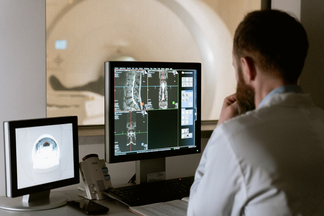
Magnetic Resonance Imaging (MRI)
MRI utilizes a powerful magnetic field and radio waves to create detailed images of the body. Unlike X-rays and CT scans, MRI does not involve the use of ionizing radiation, making it a safer option for certain populations.
MRI is highly effective in visualizing soft tissues, such as the brain, spinal cord, and joints, making it a valuable tool for detecting conditions like tumors, neurological disorders, and musculoskeletal injuries.
Ultrasound Imaging
Ultrasound imaging, also known as sonography, uses high-frequency sound waves to generate images of the body’s internal structures.
A handheld device called a transducer is moved over the skin, emitting sound waves that bounce back when they encounter different tissues. These sound waves are then converted into visual images. Ultrasound is commonly used to monitor pregnancies and examine organs such as the heart, liver, and kidneys.
Positron Emission Tomography (PET) Scan
PET scans involve the injection of a small amount of radioactive material into the body. This material accumulates in organs and tissues, emitting positrons (positively charged particles).
The collision of these positrons with electrons in the body produces gamma rays, which are detected by a specialized camera. PET scans are useful in identifying metabolic abnormalities and detecting cancerous cells.
Related Article
Visual Indicators of Health Problems in Pictures

Single-Photon Emission Computed Tomography (SPECT)
SPECT is a nuclear imaging technique that involves the injection of a radioactive substance into the bloodstream. This substance emits gamma rays, which are detected by a specialized camera.
SPECT scans are commonly used to diagnose and monitor conditions such as heart disease, brain disorders, and bone abnormalities.
Fluoroscopy
Fluoroscopy is a real-time imaging technique that uses X-rays to produce continuous images of the body. It is commonly used in procedures such as angiography (visualizing blood vessels), gastrointestinal examinations, and orthopedic surgeries.
Fluoroscopy helps guide physicians during interventions and enables them to monitor the progress of treatments in real-time.
Endoscopy
While not strictly an imaging technique, endoscopy is a valuable tool for visualizing internal structures and diagnosing health problems.
It involves the insertion of a flexible tube with a light and camera (endoscope) through an orifice or a small incision. This allows healthcare professionals to directly visualize organs and tissues, making it an essential procedure for detecting gastrointestinal disorders, respiratory conditions, and certain cancers.
Nuclear Medicine Imaging
Nuclear medicine imaging involves the administration of a small amount of radioactive material into the body. This material accumulates in specific organs or tissues, emitting gamma rays that can be detected by a specialized camera.
This imaging technique is particularly useful in identifying functional abnormalities in organs, such as the heart, liver, and thyroid, as well as detecting infections, tumors, and bone disorders.
Thermography
Thermography is a non-invasive imaging technique that detects and measures thermal patterns on the body’s surface. It uses specialized cameras to capture infrared radiation emitted by the body.
Thermographic images are analyzed for abnormal temperature variations, which can indicate inflammation, circulation problems, or certain types of cancer.
Conclusion
Imaging techniques have revolutionized the field of medical diagnostics, enabling healthcare professionals to spot and diagnose various health problems with greater precision.
From X-rays and CT scans to MRI and ultrasound imaging, each technique offers unique advantages and plays a vital role in different clinical scenarios. By leveraging these advanced imaging technologies, medical professionals can detect abnormalities early on, leading to better patient outcomes and more effective treatment plans.


