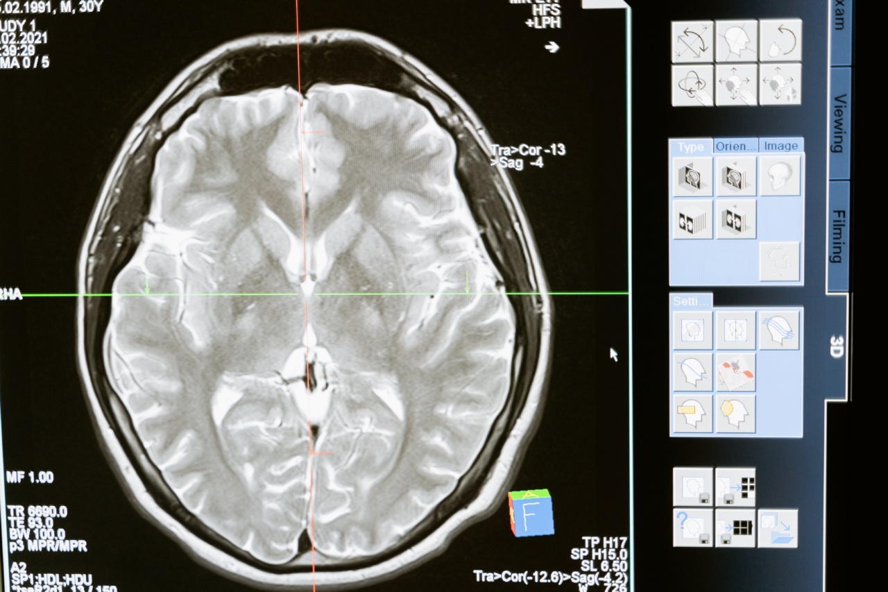
Lung cancer is a leading cause of cancer-related deaths worldwide, with a high mortality rate due to late-stage diagnosis. Early detection and accurate diagnosis are crucial for effective treatment and improved patient outcomes.
Computer-assisted analysis has emerged as a valuable tool in improving lung cancer diagnosis by aiding in the identification and characterization of suspicious findings on imaging studies. This article explores the advancements in computer-assisted analysis techniques and their impact on lung cancer diagnosis.
Current Challenges in Lung Cancer Diagnosis
Diagnosing lung cancer can be challenging due to the overlapping features of benign and malignant lung nodules. Traditional diagnostic methods, such as visual interpretation of imaging studies, are subjective and prone to inter-observer variability.
This often leads to missed diagnoses or unnecessary invasive procedures.
Computer-Aided Diagnosis (CAD) Systems
Computer-aided diagnosis (CAD) systems use advanced algorithms and artificial intelligence to support radiologists in the interpretation of medical images.
These systems can assist in the detection, classification, and quantification of lung nodules, improving diagnostic accuracy and efficiency.
Improved Nodule Detection
CAD systems can automatically detect lung nodules on chest radiographs and computed tomography (CT) scans.
By analyzing image characteristics such as size, shape, and texture, CAD algorithms can identify suspicious nodules that may have been overlooked by human observers. This enhances the sensitivity of lung cancer screening programs and facilitates early detection.
Nodule Characterization
Characterizing lung nodules as benign or malignant is crucial for determining appropriate management strategies.
CAD systems can analyze various imaging features, including nodule size, shape, density, and margins, to provide a quantitative assessment of the likelihood of malignancy. This information can help radiologists make more accurate diagnostic decisions and decrease unnecessary invasive procedures.
Integration of Clinical Data
CAD systems can integrate clinical data, such as patient demographics, medical history, and laboratory results, to further enhance lung cancer diagnosis.
By considering additional patient-specific factors, CAD algorithms can provide personalized risk assessment and treatment recommendations.
Advancements in Artificial Intelligence
The field of artificial intelligence (AI) has significantly contributed to the advancement of computer-assisted lung cancer diagnosis.
Deep learning algorithms, a subset of AI, have shown remarkable performance in image recognition and interpretation tasks. These algorithms can learn from vast amounts of annotated imaging data to improve their accuracy over time.
Automated Segmentation and Measurement
Accurate segmentation and measurement of lung nodules are essential for precise diagnosis and monitoring of treatment response. CAD systems can automatically segment lung nodules, providing accurate volumetric measurements and growth rate assessments.
This information is valuable for determining tumor progression and guiding therapeutic interventions.
Virtual Biopsy
In some cases, obtaining tissue samples through invasive procedures like biopsies may be challenging or risky. CAD systems can provide virtual biopsies by analyzing imaging data to predict the likelihood of malignancy.
This non-invasive approach reduces the need for invasive procedures, minimizing patient discomfort and complications.
Integration with Radiomics
Radiomics is an emerging field that extracts a large number of quantitative features from medical images. CAD systems can integrate radiomics analysis, combining image-based features with clinical and genetic information.
This comprehensive approach has the potential to improve lung cancer diagnosis by identifying subtle imaging biomarkers and optimizing treatment planning.
Challenges and Future Directions
Although computer-assisted analysis has shown promising results in improving lung cancer diagnosis, several challenges remain. The availability of high-quality annotated imaging data for algorithm training is essential.
Additionally, the implementation and validation of CAD systems in clinical practice require careful consideration of regulatory approval and integration with existing workflows.
Conclusion
Computer-assisted analysis has the potential to revolutionize lung cancer diagnosis by improving accuracy, efficiency, and personalized patient care.
CAD systems aid in the early detection of lung nodules, accurate characterization of malignancy risk, and guidance for treatment decisions. With further advancements in artificial intelligence and integration with radiomics, computer-assisted analysis is poised to play a crucial role in the fight against lung cancer.


