Breast ultrasound imaging is an essential diagnostic tool used to detect and evaluate abnormalities in the breast tissue.
Over the years, several technological innovations have revolutionized the field of breast ultrasound, enhancing its accuracy, efficiency, and overall patient care. In this article, we will explore some of the most significant advancements in breast ultrasound imaging.
High-Frequency Ultrasound
One of the major breakthroughs in breast ultrasound imaging is the development of high-frequency ultrasound transducers.
These transducers operate at frequencies greater than 10 MHz, allowing for better resolution and increased sensitivity in detecting subtle abnormalities in breast tissue.
Elastography
Elastography is a technique that assesses tissue stiffness or elasticity. This innovation has greatly improved the accuracy of breast ultrasound imaging by differentiating benign lesions from malignant ones.
By applying pressure to the breast tissue, elastography measures the deformation and provides valuable information regarding the stiffness of the tissue.
Automated Breast Ultrasound
Automated breast ultrasound (ABUS) is another significant advancement in breast imaging. ABUS uses a computer-controlled scanning technique that generates three-dimensional images of the entire breast.
This technique not only increases the detection rate of breast cancers, especially in women with dense breast tissue but also reduces the dependency on operator skills and variability.
Contrast-Enhanced Ultrasound
Contrast-enhanced ultrasound (CEUS) is a relatively new technique that involves the injection of a contrast agent to enhance the visibility of blood vessels, tumor vascularity, and abnormal lesions.
This innovation has proved particularly useful in characterizing focal lesions found during routine breast ultrasound examinations, improving diagnostic accuracy and reducing the need for invasive procedures.
Shear Wave Elastography
Shear wave elastography (SWE) is an advanced elastography technique that provides quantitative measurements of tissue stiffness.
Related Article
Advancements in Diagnostic Breast Ultrasound

This innovation enables accurate mapping of the tissue elasticity within a lesion, assisting in the differentiation of benign and malignant abnormalities. SWE has shown promising results in improving the specificity of breast ultrasound imaging.
Automated Lesion Detection
The development of computer-aided detection and diagnosis (CAD) software has significantly improved the efficiency and accuracy of breast ultrasound imaging.
CAD algorithms can automatically identify and classify suspicious lesions, reducing interpretation errors and assisting radiologists in making more informed diagnostic decisions.
Molecular Breast Imaging
Molecular breast imaging (MBI), also known as breast-specific gamma imaging (BSGI), is a functional imaging technique that uses a radioactive tracer to detect breast abnormalities.
MBI has proven to be highly sensitive in detecting small breast cancers, especially in women with dense breast tissue, where mammography may miss certain lesions.
Image-Guided Breast Biopsies
Breast ultrasound imaging has become an essential tool for image-guided breast biopsies.
Innovations in ultrasound technology, such as real-time imaging and improved needle visualization, have made ultrasound-guided biopsies more accurate and less invasive. These advancements allow for precise targeting of suspicious lesions, reducing the need for open surgical biopsies.
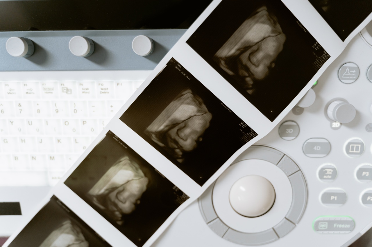
3D/4D Ultrasound
The implementation of three-dimensional (3D) and four-dimensional (4D) ultrasound in breast imaging has added another dimension to the visualization and interpretation of breast abnormalities.
3D/4D ultrasound provides volumetric data, allowing for a better assessment of lesion morphology, size, and spatial relationships within the breast tissue.
Artificial Intelligence and Deep Learning
Artificial intelligence (AI) and deep learning algorithms have shown immense potential in improving the accuracy and efficiency of breast ultrasound imaging.
By analyzing large volumes of data and patterns, AI can assist radiologists in lesion detection, classification, and risk stratification. This technology holds the promise of reducing interpretational variability and enhancing overall patient care.


