The small bowel, also known as the small intestine, plays a crucial role in the digestion and absorption of nutrients in the human body. It is located between the stomach and the large intestine and is approximately 20 feet long.
Due to its intricate structure and position, diagnosing disorders or abnormalities in the small bowel has always been a challenge for medical professionals. However, advancements in medical technology have led to the development of innovative methods for small bowel visualization and diagnosis, revolutionizing the way this part of the digestive system is examined.
1. Capsule Endoscopy
Capsule endoscopy is a non-invasive procedure that involves swallowing a small camera capsule, approximately the size of a large vitamin pill.
As the capsule moves through the digestive system, it captures high-quality images and videos of the small bowel, which can be later analyzed by a medical professional. This method allows for the visualization of the entire small bowel, providing valuable insights into any abnormalities, such as ulcers, tumors, or bleeding.
2. Double-Balloon Endoscopy
Double-balloon endoscopy is a specialized procedure that allows for the thorough examination of the small bowel. It involves the insertion of an endoscope equipped with two inflatable balloons, one at the tip and another on the side.
The balloons are inflated and deflated to advance the endoscope through the small bowel, effectively allowing for better visualization and access to the entire length of the intestine. This method is especially useful for interventions, such as biopsies or the removal of polyps.
3. Single-Balloon Enteroscopy
Similar to double-balloon endoscopy, single-balloon enteroscopy utilizes a single inflatable balloon on an endoscope to visualize and diagnose small bowel disorders.
This method is less invasive than double-balloon endoscopy but still allows for thorough examination of the small bowel. It is particularly useful in identifying and treating sources of bleeding, strictures, or inflammatory bowel disease in the small intestine.
4. Magnetic Resonance Enterography (MRE)
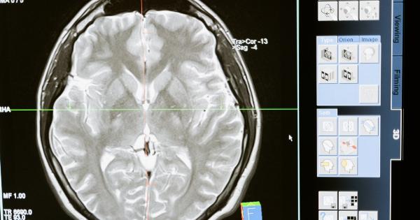
Magnetic resonance enterography (MRE) is a diagnostic imaging technique that uses magnetic resonance imaging (MRI) to create detailed images of the small intestine.
Unlike traditional X-rays or computed tomography (CT) scans, MRE does not involve the use of ionizing radiation. Instead, it utilizes a combination of magnetic fields and radio waves to generate accurate images of the small bowel. MRE is especially valuable in diagnosing Crohn’s disease, small bowel tumors, or strictures.
5. Wireless Capsule Motility
Wireless capsule motility is a method that combines capsule endoscopy with pressure sensors.
Specialized capsules equipped with pressure sensors are swallowed, and as they move through the small bowel, they measure and record the pressure changes within the intestine. This information can provide insights into the motility and transit time within the small bowel, aiding in the diagnosis of motility disorders and functional gastrointestinal issues, such as irritable bowel syndrome (IBS).
6. Small Bowel Contrast Ultrasonography
Small bowel contrast ultrasonography utilizes ultrasound technology to visualize the small bowel. Contrast agents, such as oral solutions containing microbubbles, are ingested by the patient before the procedure.
These microbubbles enhance the ultrasound images, allowing for clearer visualization of the small bowel walls and any abnormalities present. Small bowel contrast ultrasonography is a non-invasive technique that is particularly valuable in diagnosing small bowel obstruction or inflammation.
7. Virtual Chromoendoscopy
Virtual chromoendoscopy is a technique that enhances the visualization of the small bowel during endoscopy procedures.
It involves the use of specific filters or software algorithms to highlight certain features within the images captured by the endoscope. By enhancing the contrast and color saturation, virtual chromoendoscopy can aid in the identification of small bowel abnormalities, such as mucosal lesions or bleeding, that may be challenging to detect in traditional endoscopy procedures.
8. Confocal Laser Endomicroscopy
Confocal laser endomicroscopy (CLE) is a real-time imaging method that provides high-resolution microscopic images of the small bowel mucosa.
It involves the use of a special endoscope with a laser scanning system that emits a laser beam into the tissue. The reflected light is then analyzed to create detailed images of the small bowel at the cellular level. CLE can help in the detection and characterization of small bowel disorders, such as celiac disease or microscopic colitis.
9. Chromoendoscopy
Chromoendoscopy is a technique that involves the application of dyes or stains to the small bowel during endoscopy procedures. The dyes highlight specific areas or lesions, making them more visible and aiding in their identification.
Chromoendoscopy can be particularly useful in detecting and diagnosing small bowel tumors, inflammation, or precancerous lesions. The technique is safe and effective, with minimal risks or side effects.
10. Artificial Intelligence
Artificial intelligence (AI) has emerged as a powerful tool in the field of medical diagnostics, including small bowel visualization and diagnosis.
AI algorithms can analyze medical images, such as capsule endoscopy videos or MRI scans, to detect and classify abnormalities in the small bowel. These algorithms can quickly process vast amounts of data, leading to more accurate and efficient diagnoses.
AI-driven technologies have the potential to revolutionize small bowel visualization and diagnosis by improving the speed and accuracy of detection.


