A mammogram is a diagnostic screening tool used to detect breast abnormalities, such as tumors or cysts, in women. It plays a crucial role in early detection and prevention of breast cancer.
Interpreting mammogram results can be a complex process, as different findings may necessitate further tests for accurate diagnosis. In this article, we will explore the various mammogram results and discuss which additional tests may be required based on those results.
Types of Mammogram Results
When you receive your mammogram results, they may fall into one of the following categories:.
1. Normal Results:
Normal results indicate that no significant abnormalities were detected in your breast tissues during the mammogram. This is the most desired outcome, suggesting that you have a low risk of breast cancer.
However, it’s important to remember that mammograms are not foolproof, and cancer can sometimes go undetected. Regular screenings are still recommended to track any changes over time.
2. Benign (Non-cancerous) Findings:
Benign findings refer to the presence of breast conditions that are non-cancerous, such as cysts, fibroadenomas, or calcifications.
Most of these conditions do not require further testing or treatment, but it’s essential to discuss the results with your healthcare provider to determine the best course of action.
3. Suspicious Abnormalities:
If your mammogram shows suspicious abnormalities, the radiologist may recommend additional tests to gather more information and make an accurate diagnosis. Suspicious findings can include masses, asymmetries, or suspicious calcifications.
It is crucial not to panic as many suspicious findings turn out to be benign. Further testing is required to provide a definitive diagnosis.
Further Tests Based on Mammogram Results
Depending on the mammogram results, your healthcare provider may recommend one or more of the following tests:.
1. Diagnostic Mammogram:
A diagnostic mammogram is a more detailed examination performed if the screening mammogram shows areas of concern. It involves additional X-rays from different angles and focuses on specific areas of interest.
The goal is to obtain a clearer view of the breast tissue to evaluate any abnormalities more accurately.
2. Breast Ultrasound:
An ultrasound uses sound waves to produce images of the breast tissue. It is often used as a complementary test to mammography.
Ultrasound can help determine whether a suspicious mass is solid or fluid-filled, leading to a better understanding of the nature of the abnormality.
3. Breast MRI:
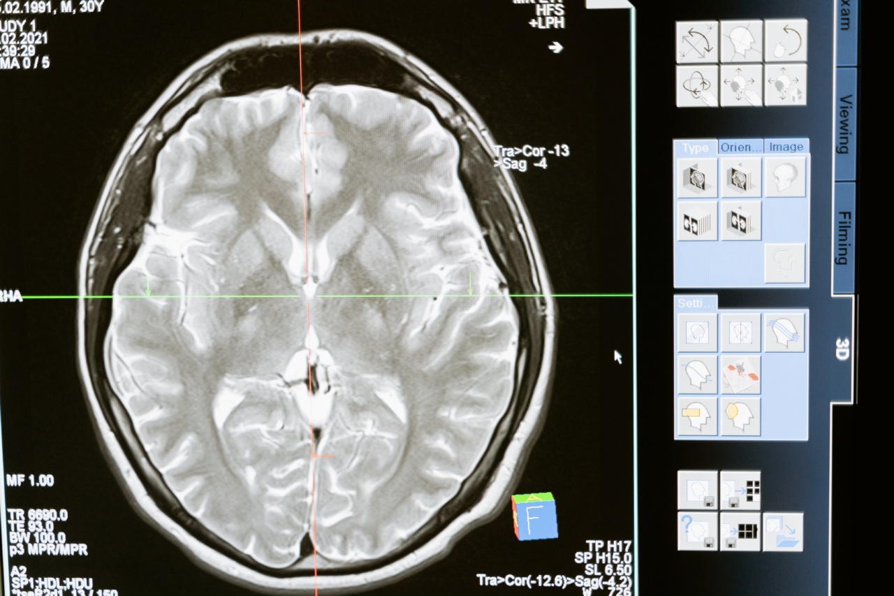
A breast magnetic resonance imaging (MRI) provides detailed images of the breast by using powerful magnets and radio waves.
It is generally recommended for individuals with a higher risk of breast cancer or when a mammogram or ultrasound does not provide a definitive diagnosis. Breast MRI is especially valuable in evaluating dense breast tissue.
4. Biopsy:
A biopsy involves the removal of a small sample of breast tissue for microscopic examination. There are several biopsy techniques, including fine-needle aspiration, core needle biopsy, and surgical biopsy.
A biopsy is the most reliable way to confirm whether a breast abnormality is cancerous or non-cancerous.
5. Ductography (Galactography):
Ductography is a specialized imaging technique that involves injecting a contrast medium into the milk ducts within the nipple. It helps visualize the inside of the ducts and identify abnormalities, such as intraductal papillomas or blocked ducts.
Ductography is usually performed when there is nipple discharge or an abnormality near the nipple.
6. Molecular Breast Imaging (MBI):
Molecular breast imaging is a nuclear medicine technique that uses a small amount of radioactive tracer to help detect breast cancer. It is often recommended for women with dense breast tissue or when other imaging tests are inconclusive.
MBI is particularly useful in identifying small breast tumors.
Monitoring Changes over Time
Depending on your age, history, and risk factors, your healthcare provider may recommend regular mammograms at specific intervals. These screenings can help monitor changes in breast tissue and detect any possible abnormalities early on.
Conclusion
Mammogram results can vary, and it’s crucial to understand the implications of different findings. Normal results bring reassurance, while benign findings require continued monitoring.
Suspicious abnormalities often warrant further tests, such as diagnostic mammograms, ultrasounds, MRIs, or biopsies, to determine whether they are cancerous. Regular mammograms, in conjunction with other screening techniques, are essential for early breast cancer detection. Always consult with your healthcare provider to determine the appropriate tests and screenings for your specific situation.


