Advancements in medical imaging technology have paved the way for a pioneering approach that allows for the precise visualization of the entire eye structure in youth, eliminating the need for pupil dilation.
This groundbreaking technique not only enhances the diagnostic process but also improves patient comfort and convenience. Traditional methods have often required pupil dilation to achieve a comprehensive view of the eye, which can cause discomfort and temporary loss of focused vision.
With this revolutionary new approach, healthcare professionals can deliver accurate diagnoses and develop effective treatment plans more efficiently than ever before.
The significance of eye structure visualization
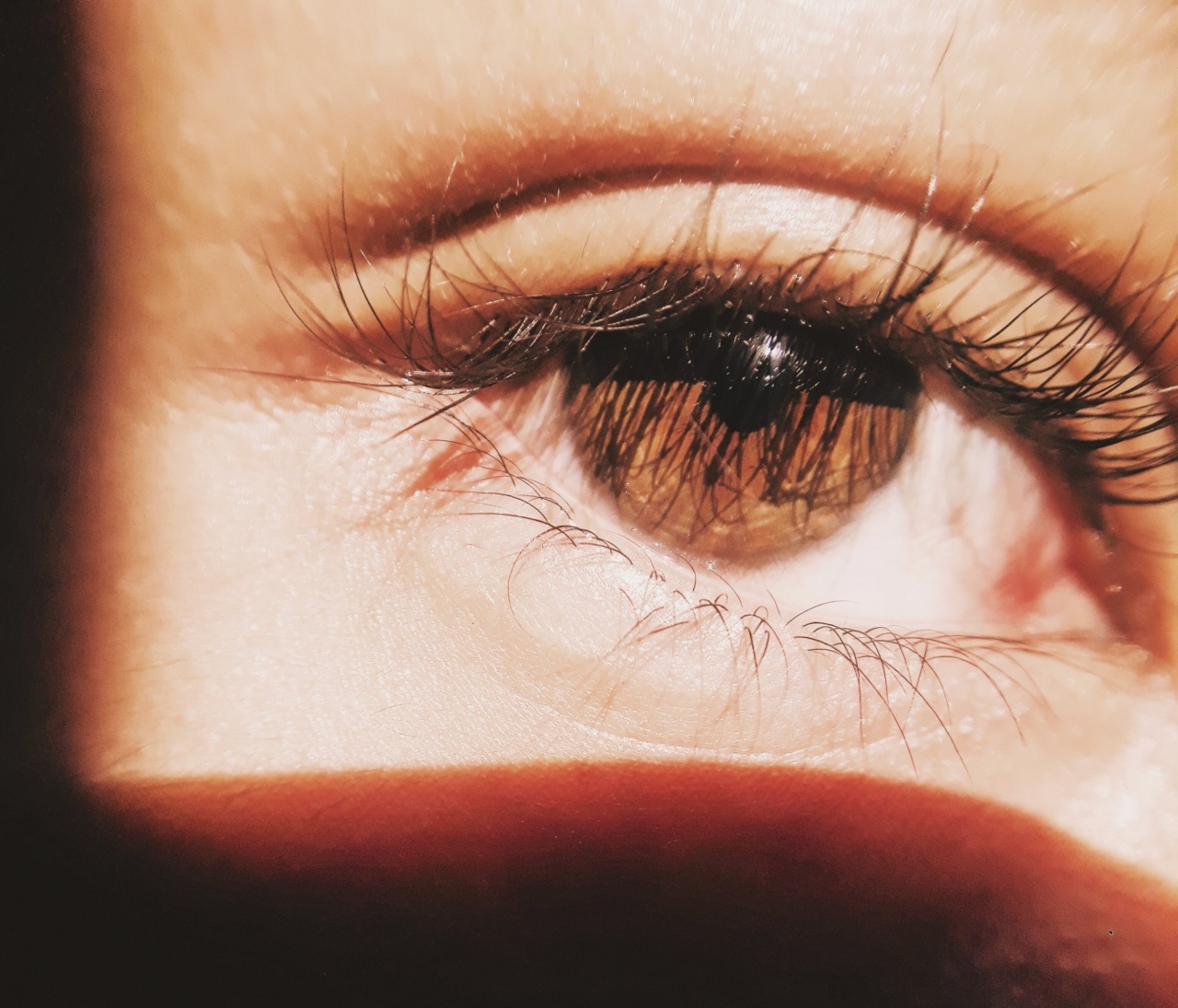
Understanding the intricate structures within the eye plays a crucial role in eye care and various fields of medicine. The eye consists of multiple components working together to enable clear vision.
Examining these structures, such as the cornea, lens, retina, and optic nerve, is essential for identifying and treating eye conditions or systemic diseases that may impact ocular health.
Traditionally, ophthalmologists have relied on pupil dilation to examine the retina and other structures at the back of the eye.
This process involves the application of dilating eye drops to widen the pupil, allowing more light to enter and providing a better view of the inner eye. However, this approach has its limitations, particularly in children and young adults.
Dilating eye drops often cause discomfort, stinging sensations, and temporary blurriness, which can be distressing for young patients. Additionally, the dilation effect can last for several hours, leading to prolonged periods of impaired near vision.
These inconveniences and side effects make it challenging to acquire comprehensive eye examinations in a timely manner.
The innovative alternative: non-dilated eye imaging
Thanks to recent advancements in imaging technology, a groundbreaking alternative to pupil dilation has emerged.
Non-dilated eye imaging techniques now make it possible to visualize the entire eye structure with exceptional clarity, regardless of pupil size. This approach utilizes specialized instruments that capture highly detailed images of the eye, allowing for accurate analysis and diagnosis without the need for dilation.
Modern imaging methods, such as optical coherence tomography (OCT), fundus photography, and ultrasound biomicroscopy, have revolutionized the way eye examinations are conducted.
These techniques provide cross-sectional images of the eye, enabling healthcare professionals to identify and evaluate conditions such as retinal disorders, glaucoma, macular degeneration, and ocular tumors.
OCT, in particular, is a non-invasive imaging technology that utilizes light waves to capture precise, high-resolution images of the retina and other ocular structures.
This painless procedure involves scanning the eye with a low-power laser beam, producing detailed cross-sectional images. By analyzing these images, ophthalmologists can assess the thickness of retinal layers, detect abnormalities, and monitor disease progression without the need for pupil dilation.
Fundus photography, another non-dilated eye imaging technique, involves capturing wide-angle images of the retina using specialized cameras.
Related Article
Advanced imaging technique captures complete eye interior sans dilation of pupil

This method allows for the visualization of the entire retina and aids in the diagnosis of conditions such as diabetic retinopathy, hypertensive retinopathy, and retinal detachments. By eliminating pupil dilation requirements, fundus photography provides a more comfortable and efficient alternative for comprehensive retinal examination.
Ultrasound biomicroscopy is an additional imaging tool that utilizes ultrasound waves to generate real-time images of the structures at the front of the eye, including the cornea, iris, and anterior chamber.
This technique is particularly useful in evaluating conditions such as angle-closure glaucoma and anterior segment tumors, offering valuable insights without any need for pupil dilation.
The benefits of non-dilated eye imaging in youth
The pioneering approach of non-dilated eye imaging offers numerous benefits, especially when considering its application in youth. Young patients, in particular, may find dilation uncomfortable and distressing.
By eliminating the need for pupil dilation, these cutting-edge imaging techniques alleviate discomfort and anxiety often associated with traditional eye exams.
Besides enhancing patient comfort, non-dilated eye imaging saves considerable time. Traditional dilation techniques can significantly prolong the duration of an eye examination, affecting both patients and healthcare providers.
By streamlining the process and eliminating dilation requirements, medical professionals can efficiently assess the eye structure, accurately diagnose conditions, and develop suitable treatment plans in a shorter timeframe.
Additionally, non-dilated eye imaging allows for more frequent examinations, enhancing preventive care and early detection of eye diseases.
When dilation is not necessary, eye health checks can be performed more regularly, enabling the timely identification and management of potential issues. This proactive approach greatly reduces the risk of complications and vision loss, particularly in youth who may be more prone to eye conditions due to their active lifestyles and exposure to various environmental factors.
Furthermore, non-dilated eye imaging offers greater convenience for patients, allowing them to resume their daily activities immediately after the examination.
The absence of post-dilation blurry vision ensures minimal disruptions, particularly for those who drive or engage in tasks that require optimal visual acuity.
Fostering collaboration and innovation
The development and adoption of non-dilated eye imaging techniques have fostered collaboration between ophthalmologists, researchers, and medical technology companies.
This collaboration has led to continual advancements in imaging accuracy, resolution, and speed, further improving the diagnostic capabilities of these techniques. The ongoing innovation in non-dilated imaging approaches promises even greater opportunities for enhanced eye care in youth and beyond.
Conclusion
The pioneering approach of non-dilated eye imaging represents a remarkable advancement in eye care, offering a more comfortable, efficient, and convenient alternative to traditional methods.
By eliminating the need for pupil dilation, healthcare professionals can now visualize the entire eye structure with exceptional clarity, enabling accurate diagnoses and treatment planning in youth without causing discomfort or temporary visual impairment. As non-dilated eye imaging techniques continue to evolve and become more widespread, the future of comprehensive eye examinations looks brighter than ever.


