Diagnostic imaging plays a crucial role in the prevention of strokes. By using advanced imaging techniques, medical professionals can identify and assess the risk factors that lead to strokes, allowing for early intervention and treatment.
In this article, we will explore the various diagnostic imaging modalities used for stroke prevention and how they contribute to patient care.
1. Computed Tomography (CT) Scan
CT scans are widely used to diagnose strokes and assess stroke-related brain damage. This imaging technique uses x-rays and computer processing to create detailed cross-sectional images of the brain.
CT scans help medical professionals identify bleeding or blockages in the blood vessels, which are common causes of strokes. With CT angiography, the visualization of blood flow is possible, aiding in the detection of stenosis or aneurysms that can increase the risk of stroke.
2. Magnetic Resonance Imaging (MRI)
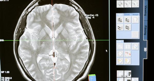
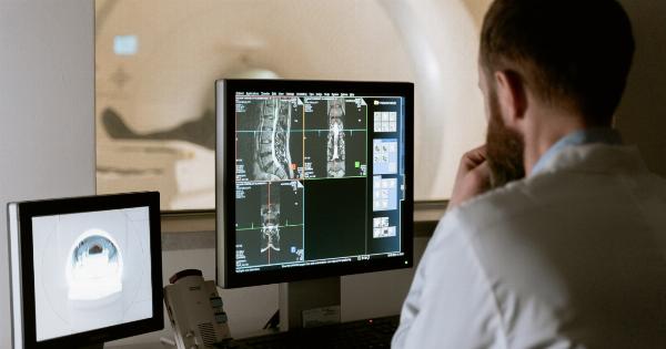
MRI scans use powerful magnets and radio waves to generate detailed images of the brain.
This non-invasive technique provides exceptional clarity and allows medical professionals to detect early signs of stroke, such as small areas of brain tissue damage. MRIs can also identify abnormal blood flow patterns, tumors, or structural abnormalities that may contribute to stroke risk.
3. Transcranial Doppler (TCD) Ultrasound
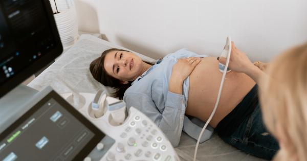
This imaging technique involves the use of ultrasound to evaluate blood flow through the brain’s blood vessels.
TCD ultrasound can detect blockages, monitor blood flow velocity, and evaluate the presence of emboli, which are small blood clots that can travel to the brain and cause strokes. TCD is particularly effective in monitoring patients who have suffered from certain types of strokes and require continuous assessment to prevent recurrent events.
4. Carotid Ultrasound
Carotid ultrasound is commonly used to assess the carotid arteries – the major blood vessels in the neck that supply blood to the brain. By using sound waves, carotid ultrasound can identify the presence of plaques or narrowing in these blood vessels.
This information is crucial in determining the risk of stroke as plaques can dislodge and cause blockages or emboli that may lead to stroke.
5. Angiography
Angiography involves the injection of contrast dye into the blood vessels to visualize their structure and blood flow.
This procedure can be performed by X-ray (conventional angiography) or by using other imaging techniques such as CT or MRI (CT angiography or MR angiography). Angiography allows medical professionals to identify any abnormalities or blockages in the blood vessels that could increase the risk of stroke.
Interventional radiologists can also employ angiography to perform minimally invasive procedures to open blockages and restore blood flow, reducing the chances of a stroke.
6. Electroencephalography (EEG)
EEG measures the electrical activity of the brain and can be used to detect abnormal patterns that might indicate a higher risk for stroke.
This non-invasive test is often employed to evaluate patients who have experienced a transient ischemic attack (TIA) or other conditions associated with an increased risk of stroke. EEG can help medical professionals understand the underlying causes of these conditions and provide appropriate treatment or preventive measures.
7. Positron Emission Tomography (PET) Scan
PET scans involve the injection of a radioactive substance into the body, which is then detected by a specialized camera.
PET scans are useful in identifying areas of reduced blood flow, metabolic abnormalities, or inflammation in the brain that may contribute to stroke risk. By detecting these abnormalities, medical professionals can formulate tailored treatment plans to prevent strokes and improve patient outcomes.
8. Magnetic Resonance Angiography (MRA)
MRA uses magnetic resonance imaging to visualize blood vessels and blood flow within the brain. This technique provides detailed images without the need for contrast dye injections.
MRA can identify narrowed or blocked arteries, identify aneurysms, and help medical professionals assess the risk of stroke. Additionally, it can detect abnormalities in blood vessels that may require surgical intervention to prevent stroke.
9. Calcium Scoring
Calcium scoring is a specialized form of CT scan that evaluates the amount of calcified plaque in the coronary arteries.
While not directly related to stroke prevention, this test helps determine a patient’s overall cardiovascular health and risk factors. As strokes often result from underlying cardiovascular conditions, such as atherosclerosis, calcium scoring provides valuable insights into the patient’s risk of stroke and prompts the initiation of appropriate preventive measures.
10. SPECT Brain Perfusion Imaging
Single-photon emission computed tomography (SPECT) measures blood flow and activity in the brain. By injecting a radioactive substance into the bloodstream, SPECT scans provide information about brain perfusion, metabolism, and neuronal activity.
This imaging technique aids in the identification of regions with inadequate blood flow, allowing medical professionals to understand the physiological changes associated with strokes and their potential causes.


