Stroke is a leading cause of death and disability worldwide, with millions of people affected each year. Preventive measures for stroke can make a significant impact on reducing the incidence of this life-threatening condition.
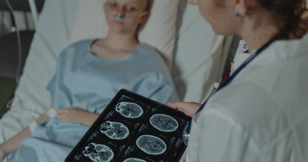
Imaging techniques play a crucial role in stroke prevention, allowing clinicians to detect and monitor risk factors and take appropriate measures to prevent the occurrence of stroke. In this article, we will discuss the various imaging techniques used for stroke prevention and the preventive measures that can be taken based on the results obtained.
Types of Imaging Techniques
Imaging techniques used in stroke prevention can be broadly classified into two categories: diagnostic and screening.
Diagnostic Imaging Techniques
Diagnostic imaging techniques are used to diagnose the presence of risk factors or the occurrence of a stroke. The following are the most common diagnostic imaging techniques used for stroke prevention:.
Magnetic Resonance Imaging (MRI)
MRI is a non-invasive imaging technique that uses strong magnetic fields and radio waves to produce detailed images of the brain.
MRI can detect the presence of risk factors such as high blood pressure, high cholesterol, and diabetes, which are known to increase the risk of stroke. MRI is also used to diagnose the occurrence of a stroke and to determine the extent of brain damage caused by a stroke.
Computed Tomography (CT) Scan
CT scan is a non-invasive imaging technique that uses X-rays to produce detailed images of the brain. CT scan can detect the presence of risk factors such as blood clots, aneurysms, and other abnormalities that increase the risk of stroke.
CT scan is also used to diagnose the occurrence of a stroke and to determine the extent of brain damage caused by a stroke.
Doppler Ultrasound
Doppler ultrasound is a non-invasive imaging technique that uses high-frequency sound waves to produce images of blood flow in the arteries and veins of the neck and head.
Doppler ultrasound can detect the presence of atherosclerosis, a condition where the arteries become narrowed due to the buildup of plaque, which is a major risk factor for stroke.
Screening Imaging Techniques
Screening imaging techniques are used to identify individuals who are at risk of stroke but do not show any symptoms. The following are the most common screening imaging techniques used for stroke prevention:.
Carotid Ultrasound
Carotid ultrasound is a non-invasive imaging technique that uses high-frequency sound waves to produce images of the carotid arteries, which supply blood to the brain.
Carotid ultrasound can detect the presence of atherosclerosis, which is a major risk factor for stroke.
Magnetic Resonance Angiography (MRA)
MRA is a non-invasive imaging technique that uses strong magnetic fields and radio waves to produce detailed images of the blood vessels in the brain.
MRA can detect the presence of abnormalities in the blood vessels, such as aneurysms or arteriovenous malformations (AVMs), which are known to increase the risk of stroke.
Computed Tomography Angiography (CTA)
CTA is a non-invasive imaging technique that uses X-rays to produce detailed images of the blood vessels in the brain.
CTA can detect the presence of abnormalities in the blood vessels, such as aneurysms or AVMs, which are known to increase the risk of stroke.
Preventive Measures Based on Imaging Results
Preventive measures for stroke depend on the individual’s risk factors and the results of imaging studies. The following are the most common preventive measures taken based on imaging results:.
Reduce High Blood Pressure
High blood pressure is a major risk factor for stroke, and reducing it can significantly reduce the risk of stroke.
If imaging studies reveal the presence of high blood pressure, lifestyle changes such as diet modifications, exercise, and weight loss, and medications such as ACE inhibitors or angiotensin receptor blockers may be prescribed to control blood pressure.
Control High Cholesterol
High cholesterol is another major risk factor for stroke, and controlling it can significantly reduce the risk of stroke.
If imaging studies reveal the presence of high cholesterol, lifestyle changes such as diet modifications, exercise, and weight loss, and medications such as statins may be prescribed to control cholesterol levels.
Treat Diabetes
People with diabetes are at a higher risk of stroke, and controlling diabetes can significantly reduce the risk of stroke.
If imaging studies reveal the presence of diabetes, lifestyle changes such as diet modifications, exercise, and weight loss, and medications such as insulin or oral hypoglycemics may be prescribed to control blood sugar.
Prevent Blood Clots
Blood clots are a major cause of stroke, and preventing them can significantly reduce the risk of stroke.
If imaging studies reveal the presence of blood clots, medications such as aspirin, anticoagulants, or antiplatelets may be prescribed to prevent blood clots.
Surgical Intervention
In some cases, surgical intervention may be required to prevent stroke. For example, if imaging studies reveal the presence of an aneurysm or AVM, surgery may be required to repair or remove the abnormality and reduce the risk of stroke.
Conclusion
Preventing stroke is a critical aspect of healthcare, and imaging techniques play a crucial role in stroke prevention by allowing clinicians to detect and monitor risk factors and take appropriate measures to prevent the occurrence of stroke.
The preventive measures taken depend on individual risk factors and imaging results and may include lifestyle changes, medications, or surgical intervention. By taking preventive measures based on imaging results, we can reduce the incidence of this life-threatening condition and improve the health and well-being of millions of people around the world.


