Prostate cancer is one of the most common types of cancer in men, affecting millions of individuals worldwide. Early and accurate diagnosis is crucial for successful treatment and improved patient outcomes.
Traditional diagnostic methods, such as digital rectal examination and prostate-specific antigen (PSA) tests, have limitations in terms of accuracy and reliability. However, with advancements in medical imaging technology, polyparametric magnetic resonance imaging (MRI) has emerged as a revolutionary tool for the diagnosis of prostate cancer.
What is Polyparametric Magnetic Resonance?
Polyparametric magnetic resonance combines multiple imaging techniques to obtain a comprehensive evaluation of the prostate gland.
The techniques involved in polyparametric MRI include T2-weighted imaging, diffusion-weighted imaging (DWI), dynamic contrast-enhanced imaging (DCE-MRI), and magnetic resonance spectroscopy (MRS).
T2-Weighted Imaging
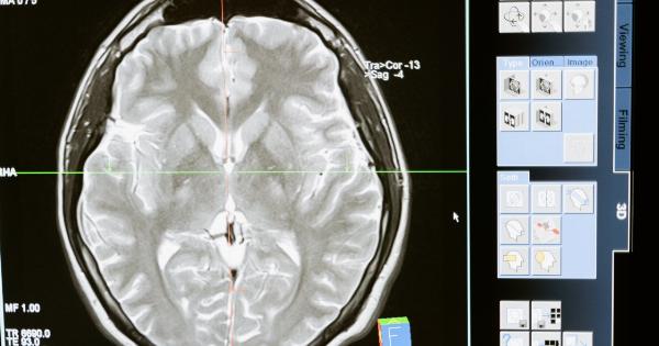
T2-weighted imaging is the most basic component of polyparametric MRI. It generates high-resolution images of the prostate gland and surrounding structures, allowing for the detection of abnormal areas.
Tumors often appear as dark regions on T2-weighted images.
Diffusion-Weighted Imaging (DWI)
DWI evaluates the diffusion of water molecules within prostate tissues. Cancerous tissues tend to have restricted water diffusion compared to healthy tissues.
DWI provides valuable information about tissue cellularity and can aid in the identification and assessment of prostate tumors.
Dynamic Contrast-Enhanced Imaging (DCE-MRI)
DCE-MRI involves the injection of a contrast agent to evaluate the blood flow and vascular permeability within the prostate gland. Cancerous tissues often exhibit increased blood flow and enhanced contrast agent uptake.
DCE-MRI helps in distinguishing between malignant and benign lesions and provides information about the aggressiveness and stage of the cancer.
Magnetic Resonance Spectroscopy (MRS)
MRS measures the concentration of specific metabolites within prostate tissues. It can help in differentiating between cancerous and non-cancerous tissues based on the metabolic profiles.
MRS acts as a complementary tool to other imaging techniques, enhancing the accuracy of prostate cancer diagnosis.
Advantages of Polyparametric MRI for Prostate Cancer Diagnosis
Polyparametric MRI offers numerous advantages over conventional diagnostic methods for prostate cancer. Some of the notable advantages include:.
1. Improved Sensitivity and Specificity
The combination of multiple imaging techniques in polyparametric MRI improves the sensitivity and specificity of prostate cancer detection.
It allows for a more accurate localization and characterization of tumors, minimizing false-positive and false-negative results.
2. Non-Invasiveness
Polyparametric MRI is a non-invasive diagnostic technique that does not require the use of ionizing radiation or invasive procedures. This reduces patient discomfort and eliminates the risk of complications associated with invasive diagnostic methods.
3. Early Detection and Staging
Polyparametric MRI can detect prostate cancers at an early stage when they are small and confined to the gland. It also provides valuable information about the extent of tumor spread, allowing for accurate staging of prostate cancer.
4. Guided Biopsies
Polyparametric MRI can be used to guide prostate biopsies, ensuring accurate sampling of suspicious regions. This reduces the number of unnecessary biopsies and increases the chances of detecting clinically significant tumors.
5. Treatment Planning
Polyparametric MRI assists in treatment planning by providing detailed information about tumor location, size, and aggressiveness.
This helps in selecting the most appropriate treatment approach, such as surgery, radiation therapy, or active surveillance.
Limitations and Challenges
While polyparametric MRI is a revolutionary tool for prostate cancer diagnosis, it is not without limitations and challenges.
1. Cost and Availability
Polyparametric MRI is relatively expensive compared to conventional diagnostic methods, which can limit its accessibility in certain healthcare settings.
Additionally, specialized equipment and expertise are required for performing and interpreting polyparametric MRI scans.
2. False-Positive and False-Negative Results
Although polyparametric MRI improves the accuracy of prostate cancer diagnosis, there is still a possibility of false-positive and false-negative results.
Some benign conditions or inflammation can mimic cancerous lesions, leading to unnecessary biopsies or missed diagnoses.
3. Limitations in Assessing Lymph Node Involvement
Polyparametric MRI has limitations in accurately assessing lymph node involvement in prostate cancer. However, this can be complemented by other imaging techniques or through the use of targeted molecular imaging.
Future Directions and Conclusion
Polyparametric magnetic resonance imaging has revolutionized the diagnosis of prostate cancer, offering improved sensitivity, specificity, and non-invasiveness.
Ongoing research aims to further enhance the accuracy and accessibility of this diagnostic tool. With advancements in imaging technology, such as artificial intelligence-based image analysis and functional imaging techniques, the future of prostate cancer diagnosis looks promising.


