Parkinson’s disease is a neurodegenerative disorder that affects millions of people worldwide. It is characterized by a lack of dopamine production in the brain, leading to symptoms such as tremors, stiffness, and difficulty with movement.
While there is currently no cure for Parkinson’s, early detection and intervention can greatly improve the quality of life for those living with the disease.
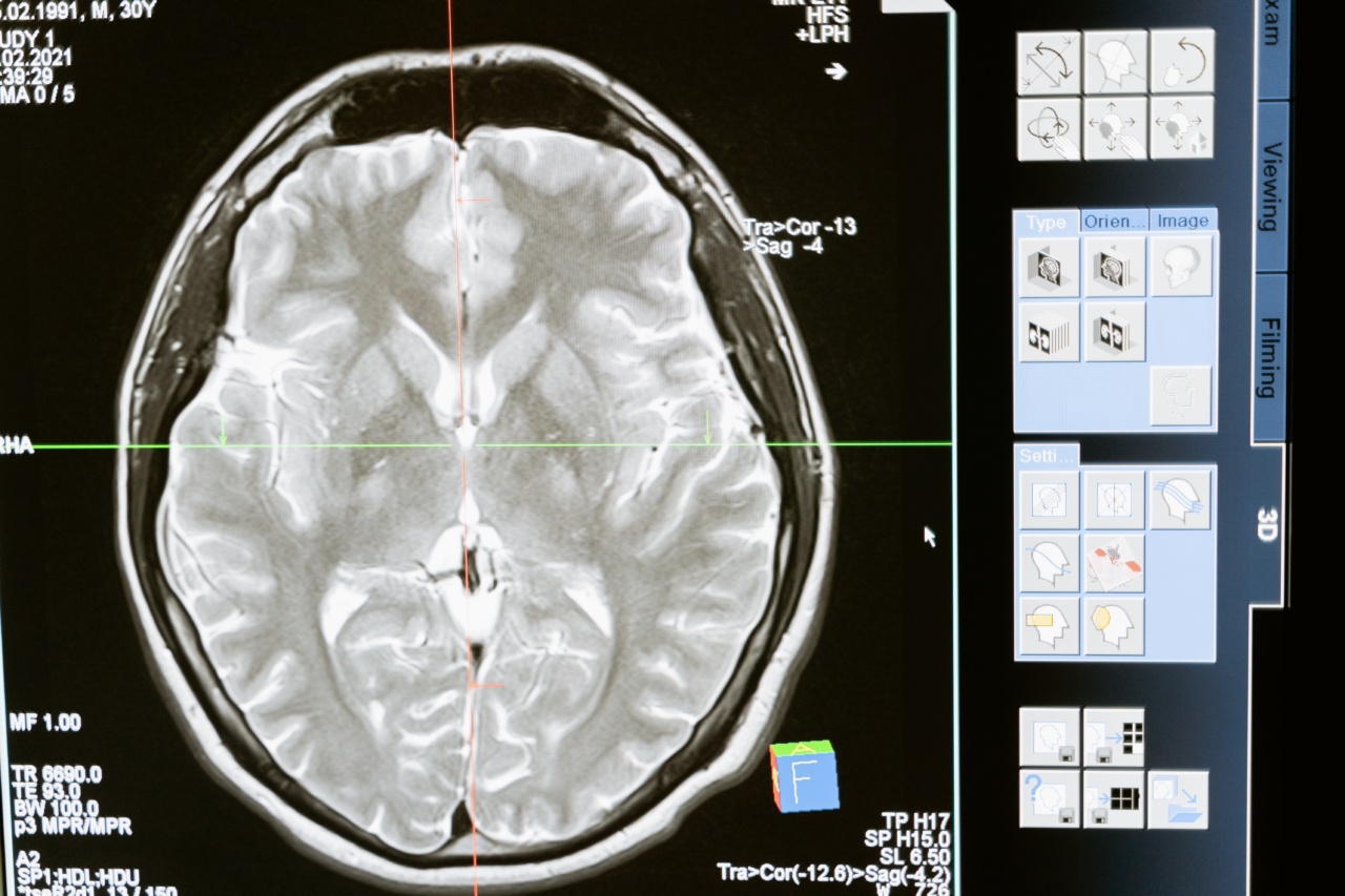
Recent advancements in brain scanning technology have made it possible to identify Parkinson’s disease before any symptoms manifest.
This groundbreaking development has the potential to revolutionize the way Parkinson’s is diagnosed and treated, providing hope for patients and their families.
The Role of Brain Scanning in Parkinson’s Diagnosis
Traditionally, the diagnosis of Parkinson’s disease has relied on the observation of physical symptoms during a clinical examination.
However, by the time these symptoms become apparent, a significant amount of dopamine-producing cells in the brain have already been damaged or lost.
Special brain scanning techniques, such as functional MRI (fMRI) and positron emission tomography (PET), offer a non-invasive way to visualize changes in the brain that occur before symptoms arise.
These scans can detect abnormal activity in specific regions of the brain associated with dopamine production, providing early indicators of Parkinson’s disease.
The Promise of fMRI in Parkinson’s Detection
Functional MRI (fMRI) is a type of brain imaging that measures changes in blood flow and oxygen levels in the brain. These changes are correlated with neuronal activity, allowing researchers to map brain activity in real-time.
In the case of Parkinson’s disease, fMRI scans can reveal abnormalities in the basal ganglia and other areas that are affected by dopamine loss.
A study conducted at the University of California, San Francisco, used fMRI to track brain changes in individuals at high risk of developing Parkinson’s disease due to genetic factors or other known risk factors.
The researchers found distinct patterns of altered activity in the brain even before the onset of motor symptoms.
By identifying these early changes, doctors can intervene with targeted interventions to slow down or even prevent the progression of the disease.
This early detection could significantly enhance treatment outcomes and quality of life for individuals with Parkinson’s.
The Role of PET Scans in Parkinson’s Detection
Positron emission tomography (PET) is another powerful tool in detecting Parkinson’s disease before symptoms appear.
Related Article
New Brain Scanning Technique Can Diagnose Parkinson’s Early

Unlike fMRI, which measures brain activity indirectly, PET scans directly identify changes in specific molecules and receptors in the brain.
In the case of Parkinson’s disease, PET scans can evaluate the levels of dopamine and other neurotransmitters in the brain.
A study published in the Journal of Nuclear Medicine found that individuals with a specific genetic mutation associated with Parkinson’s disease had reduced dopamine transporters even before the onset of symptoms.
Early detection of these changes allows for timely intervention and treatment, potentially slowing the progression of the disease and improving patient outcomes.
Challenges and Future Directions
While the potential for early detection of Parkinson’s disease through brain scanning is promising, several challenges remain to be addressed.
One major challenge is the cost and accessibility of these specialized brain scanning techniques. Currently, fMRI and PET scans are expensive and not widely available in all healthcare settings.
Increasing accessibility and reducing costs are crucial for widespread adoption of these technologies in Parkinson’s disease diagnosis.
Another challenge lies in distinguishing the early brain changes associated with Parkinson’s disease from other neurological conditions.
Many symptoms of Parkinson’s, such as tremors and movement difficulties, can also be present in other disorders. Further research is needed to develop specific biomarkers that can differentiate Parkinson’s disease from other conditions with similar symptoms.
Despite these challenges, the potential of special brain scanning in identifying Parkinson’s disease before symptoms develop cannot be overstated.
Early detection would not only allow for timely intervention but also enable researchers to study the disease progression more effectively and develop novel therapeutic approaches.
Conclusion
In conclusion, recent advancements in brain scanning technology have unlocked the potential to identify Parkinson’s disease before any physical symptoms become apparent.
Techniques such as fMRI and PET scans offer a non-invasive way to detect early brain changes associated with Parkinson’s, enabling early intervention and improving patient outcomes.
While there are challenges to overcome in terms of accessibility and differentiation from other conditions, the promise of early detection in Parkinson’s disease holds great hope for those affected by the condition.
With further research and advancements in brain scanning technology, we may be able to detect and treat Parkinson’s disease at its earliest stages, changing the lives of millions of people worldwide.


