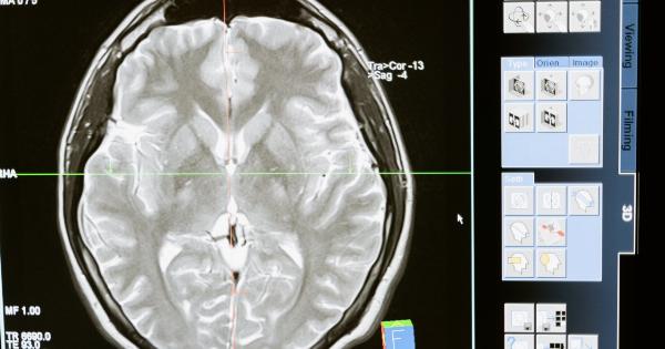
Medical imaging plays a vital role in the diagnosis and treatment of various illnesses, and X-rays are one the oldest and most widely used forms of medical imaging.
However, there has been a long-standing concern about the potential harm of radiation exposure from such imaging techniques, and its possible association with cancer. In this article, we will investigate the impact of radiation from X-rays and axial on cancer.
X-rays and Radiation
X-rays are a form of electromagnetic radiation that can penetrate tissues and bones to create images that help diagnose and treat medical conditions.
It is a non-invasive and painless procedure that can detect broken bones, lung infections, dental problems, and tumors, among other things. However, when X-rays pass through the body, they can cause ionization, which involves the transfer of energy to atoms and molecules in living cells. This ionizing radiation can lead to genetic mutations, cellular damage, and eventually, cancer.
Radiation Doses from X-rays and Axial
The radiation dose from X-rays and axial imaging varies depending on the type of procedure. It is measured in millisieverts (mSv), which is an estimate of the amount of ionizing radiation that a patient receives.
According to the United States Nuclear Regulatory Commission, the average American receives about 3mSv of radiation per year from natural sources such as cosmic rays and radon. Diagnostic X-rays expose patients to typically low levels of radiation, which range from 0.01mSv for dental X-rays to 10mSv for computed tomography (CT) scans, depending on the body area being examined.
Some complex procedures such as cardiac CT scans can deliver up to 20mSv.
Cancer Risk from X-rays and Axial
The risk from radiation exposure on cancer varies depending on the dose, the age of exposure, and the type of radiation.
Epidemiological studies on radiation workers and atomic bomb survivors have established that there is a linear, no-threshold relationship between radiation exposure and cancer risk, that is, even a small dose of radiation can increase the risk of cancer. The excess relative risk (ERR) per unit dose of radiation, which is the ratio of the increase in cancer incidence to the dose of radiation received, is estimated to be 0.05 per mSv for solid cancers and 0.15 per mSv for leukemia.
Similarly, studies on children exposed to CT scans have shown an increased risk of cancer, particularly in organs that were directly in the path of the X-rays.
Minimizing Radiation Exposure
Although the risk of cancer from X-rays and axial imaging is small, it is still important to minimize exposure to ionizing radiation whenever possible.
Radiologists and healthcare practitioners can implement several measures to help reduce the risk of cancer:.
Conclusion
The use of X-rays and axial imaging techniques has revolutionized medical diagnosis and treatment, but also presents a potential risk of cancer from radiation exposure.
It is therefore important to balance the benefits of medical imaging with the risks of radiation, by implementing appropriate radiation safety measures and using imaging alternatives whenever possible. By doing so, healthcare practitioners can ensure that their patients receive optimal care while minimizing the risk of radiation-induced cancer.


