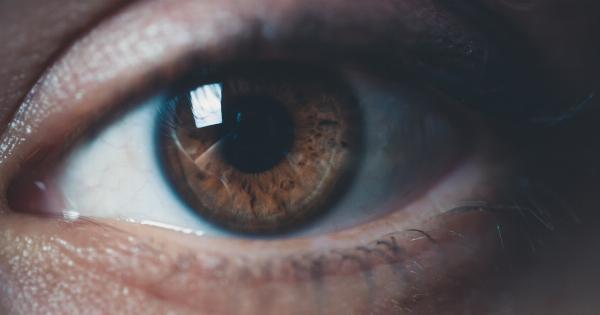
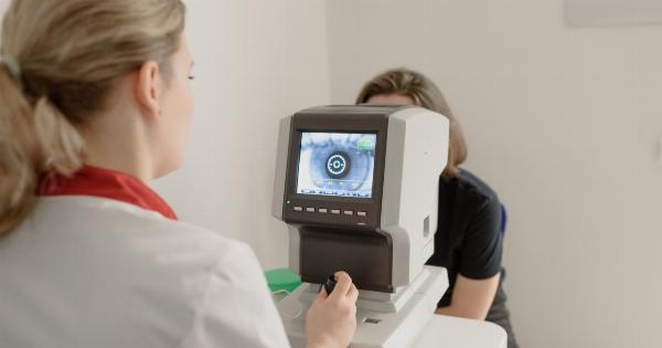
Retinal detachment occurs when the thin lining at the back of your eye, known as the retina, peels away from its normal position. This condition can potentially lead to vision loss or blindness if not promptly treated.
In this article, we will explore the top causes and treatments for retinal detachment.
Understanding the Retina
The retina is a crucial part of the eye that converts light into electrical signals, allowing us to see. It is essentially a delicate layer of tissue located at the back of the eye.
When light enters the eye, it passes through the lens and focuses onto the retina, triggering a series of chemical reactions that enable vision.
Causes of Retinal Detachment
Retinal detachment can occur due to various reasons. The common causes include:.
- Age-related degeneration: As we age, the vitreous gel inside the eye may shrink or become more liquid, leading to an increased risk of retinal detachment.
- Eye injury: Trauma or injury to the eye can cause retinal detachment.
- Family history: If you have a family history of retinal detachment, your risk of developing the condition may be higher.
- Thin or weak spots: Some people may have thin or weak areas in their retina, making them more susceptible to detachment.
- Previous eye surgery: Individuals who have undergone previous eye surgeries, such as cataract removal, are at increased risk.
- Medical conditions: Certain medical conditions such as diabetes, nearsightedness, and inflammatory disorders can increase the risk of retinal detachment.
Common Symptoms of Retinal Detachment
Retinal detachment may cause various symptoms, including:.
- Floaters: Seeing small, dark spots or specks drifting across your field of vision.
- Flashes of light: Noticing sudden flashes of light, similar to lightning bolts.
- Partial vision loss: Experiencing a dark curtain or shadow suddenly covering part of your visual field.
- Blurry or distorted vision: Seeing objects as wavy, crooked, or blurred.
- Decreased central vision: Experiencing a reduction in sharpness and clarity of the central part of your vision.
Treatments for Retinal Detachment
Retinal detachment is considered a medical emergency, and prompt treatment is crucial to prevent permanent vision loss. The primary goal of treatment is to reattach the retina and restore its proper function.
The suitable treatment option depends on the type, extent, and location of retinal detachment. Common treatments include:.
Laser Photocoagulation (Thermal Photocoagulation)
During laser photocoagulation, a high-energy laser is used to seal the tears or holes in the detached retina. The laser creates burns around the area, causing scar tissue to form, which then helps reattach the retina to the underlying tissues.
This procedure is typically performed in an eye doctor’s office and does not require surgery.
Cryopexy
Cryopexy involves using a freezing probe to freeze the area around the retinal tear or hole. This freezing process creates a scar, effectively sealing the retina and preventing further detachment.
Similar to laser photocoagulation, cryopexy can be performed in an office setting without the need for surgery.
Scleral Buckling
Scleral buckling is a surgical procedure that involves the placement of a silicone or silicone sponge-like band around the eye to support the detached retina. This band or buckle pushes the wall of the eye toward the retina, helping to reattach it.
Scleral buckling is often performed in combination with other procedures, such as laser therapy or cryopexy.
Vitrectomy
Vitrectomy is a surgical technique used to treat complex or severe cases of retinal detachment. During this procedure, the vitreous gel inside the eye is removed, and the detached retina is carefully reattached using specialized instruments.
After reattachment, the eye is typically filled with a gas or silicone oil bubble to help keep the retina in place during healing.
Pneumatic Retinopexy
Pneumatic retinopexy is a non-invasive procedure performed in an office setting. It involves injecting a small gas bubble into the vitreous cavity of the eye.
The gas bubble helps push the detached retina against the wall of the eye, allowing the tear or hole to seal. Laser therapy or cryopexy is often used with pneumatic retinopexy to permanently seal the tear or hole.
Seek Immediate Medical Attention
If you experience any symptoms of retinal detachment, it is crucial to seek immediate medical attention. Prompt treatment increases the chances of successfully reattaching the retina and preserving vision.


