Medical imaging has revolutionized the field of ophthalmology by providing valuable insights into the diagnosis and treatment of vision disorders.
With the ability to capture detailed images of the eye, medical imaging techniques such as optical coherence tomography (OCT), fundus photography, and magnetic resonance imaging (MRI) have become indispensable tools for ophthalmologists. In this article, we will explore how medical imaging is used to diagnose a wide range of vision disorders.
Optical Coherence Tomography (OCT)
One of the most commonly used imaging techniques in ophthalmology is optical coherence tomography (OCT). This non-invasive imaging modality allows ophthalmologists to obtain high-resolution, cross-sectional images of the retina.
By using light waves to create detailed images of the different layers of the retina, OCT provides insights into various eye conditions, including age-related macular degeneration (AMD), diabetic retinopathy, and glaucoma.
Fundus Photography
Fundus photography is another widely used imaging method in ophthalmology. It involves capturing photographs of the back of the eye, including the retina, optic disc, macula, and blood vessels.
These images assist ophthalmologists in diagnosing and managing conditions such as retinal detachment, hypertensive retinopathy, and optic nerve diseases. Fundus photography is particularly useful for monitoring changes in the eye over time and assessing the effectiveness of treatment.
Magnetic Resonance Imaging (MRI)
While OCT and fundus photography focus on the structures of the eye, magnetic resonance imaging (MRI) provides a broader view of the head and brain.
MRI can be instrumental in diagnosing vision disorders that originate from the brain, such as tumors, strokes, or multiple sclerosis. By capturing detailed images of the brain and its surrounding structures, MRI helps ophthalmologists determine the causes of visual impairment and plan appropriate treatment strategies.
Corneal Topography
Corneal topography is a specialized technique that maps the curvature and thickness of the cornea, the transparent front part of the eye.
By analyzing corneal topography images, ophthalmologists can diagnose and monitor conditions such as keratoconus, astigmatism, and corneal dystrophies. This information is crucial for determining the best course of treatment, such as corneal transplantation or the use of contact lenses.
Ultrasound Imaging
In some cases, especially when the view of the eye is obstructed, ultrasound imaging can be used to visualize the internal structures of the eye. This technique utilizes sound waves to create images of the eye and its surrounding tissues.
Ultrasound imaging is particularly valuable in diagnosing conditions like orbital tumors, foreign bodies, and intraocular hemorrhages. It can also aid in the evaluation of the eye during pregnancy or when the eye is injured.
Related Article
Vision impairments: Diagnostic images for common diseases

Fluorescein Angiography
Fluorescein angiography is a diagnostic tool used to assess the blood flow in the retina and detect abnormalities in the blood vessels.
During this procedure, a dye called fluorescein is injected into a vein, and a series of photographs are taken as the dye circulates through the blood vessels of the retina. This imaging technique is primarily used for evaluating conditions such as diabetic retinopathy, macular degeneration, and vascular occlusions.
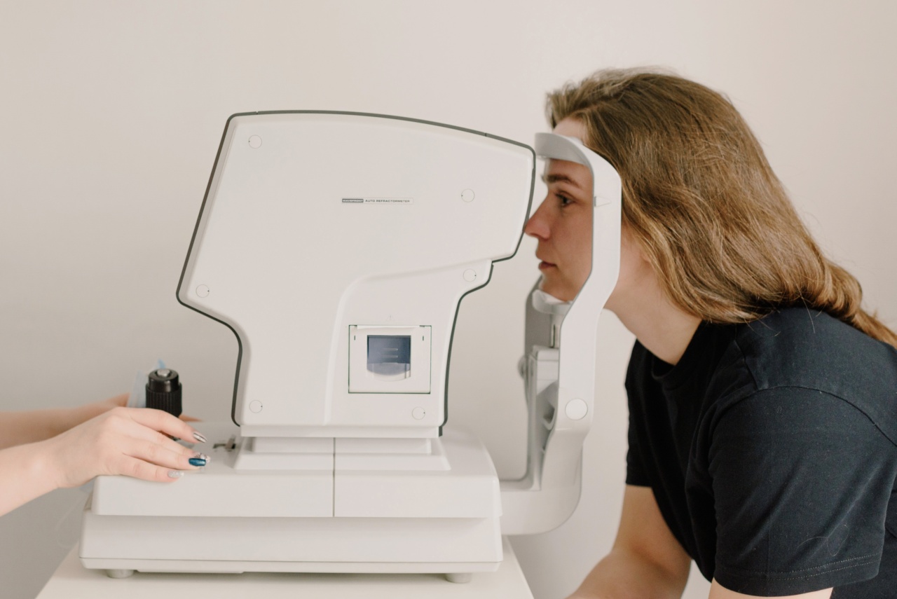
Optical Biometry
Optical biometry is a technique used to measure the dimensions of the eye, especially for determining the appropriate intraocular lens power in cataract surgery.
It utilizes optical imaging to obtain measurements such as axial length, corneal curvature, and anterior chamber depth. These measurements play a critical role in calculating the power of the artificial lens required to restore clear vision post-cataract surgery.
Electroretinography (ERG)
Electroretinography (ERG) is an electrophysiological technique used to assess the function of the retina. During an ERG test, electrodes are placed on the surface of the eye, and the retina’s response to light stimulation is recorded.
By measuring the electrical signals generated by the retina, ophthalmologists can evaluate the health and functionality of retinal cells. ERG is particularly useful in diagnosing inherited retinal diseases, like retinitis pigmentosa.
Scanning Laser Ophthalmoscopy (SLO)
Scanning laser ophthalmoscopy (SLO) is an imaging technique that provides high-resolution, three-dimensional images of the retina. It uses a laser beam to scan the retina and create detailed images of its structures.
SLO allows ophthalmologists to visualize subtle changes in the retina and is especially valuable in the early detection and monitoring of diseases like age-related macular degeneration and diabetic retinopathy.
Confocal Scanning Laser Tomography (CSLT)
Confocal scanning laser tomography (CSLT) is a powerful imaging technique used to evaluate the optic nerve head and the surrounding nerve fibers.
By providing detailed, cross-sectional images, CSLT assists in the assessment of conditions like glaucoma and optic nerve damage caused by various pathologies. It helps ophthalmologists measure the thickness of the retinal nerve fiber layer, which can indicate the progression of these diseases over time.
Conclusion
Medical imaging plays a central role in the diagnosis and management of vision disorders. From OCT and fundus photography to MRI and ultrasound imaging, these techniques provide invaluable information about the structures and functions of the eye.
By combining medical imaging with clinical expertise, ophthalmologists can accurately diagnose various vision disorders and develop effective treatment plans, ultimately improving the vision and quality of life for countless individuals.


