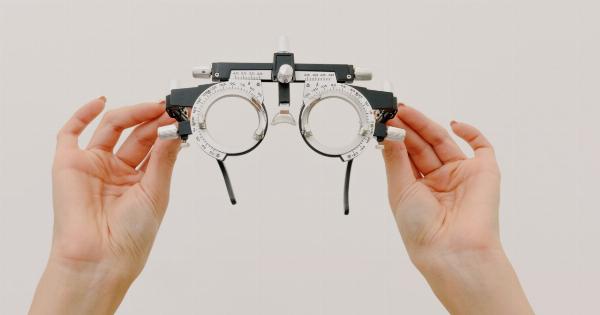
Vision impairments are medical conditions that affect the ability of an individual to see clearly. This can be caused by several underlying conditions, and each of these conditions requires a different diagnostic approach.
In this article, we will be discussing the diagnostic images used for some of the most common vision impairments.
Age-Related Macular Degeneration
Age-related macular degeneration (AMD) is a common condition that affects the eye’s macula. This condition is usually seen in elderly individuals and leads to a gradual loss of central vision.
Diagnostic imaging techniques used in the diagnosis of AMD include:.
Optical Coherence Tomography (OCT)
OCT is a non-invasive diagnostic technique that uses light waves to create cross-sectional images of the retina. This technique helps in the diagnosis of AMD by identifying areas of the macula that are thinning or developing fluid accumulation.
Fluorescein Angiography (FA)
FA is another diagnostic technique used in the diagnosis of AMD. This technique involves administering a dye into the vein of the patient’s arm, which then travels to the eye.
A specialized camera is used to take pictures of the retina as the dye flows through the retinal blood vessels. The images obtained can reveal areas where blood vessels are leaking, which is an indicator of AMD.
Glaucoma
Glaucoma is an eye disease that damages the optic nerve and can lead to vision loss. This condition is usually caused by high intraocular pressure. Diagnostic imaging techniques used in the diagnosis of glaucoma include:.
Visual Field Testing
Visual field testing measures the range of vision of an individual and can help detect areas of the visual field that are affected by glaucoma. This test is usually done with the help of a machine that projects a series of lights onto a screen.
Optical Coherence Tomography (OCT)
OCT can also be used in the diagnosis of glaucoma. In this case, OCT images are used to evaluate the thickness of the retinal nerve fiber layer, which is an indicator of glaucoma.
Diabetic Retinopathy
Diabetic retinopathy is a complication of diabetes that affects the blood vessels in the retina. This condition can lead to vision loss and, in severe cases, can cause blindness.
Diagnostic imaging techniques used in the diagnosis of diabetic retinopathy include:.
Fluorescein Angiography (FA)
FA can be used in the diagnosis of diabetic retinopathy by detecting areas of the retina where blood vessels are blocked or leaking. These areas of blood vessel damage can lead to the development of diabetic retinopathy.
Optical Coherence Tomography (OCT)
Similar to other vision impairments, OCT can be used in the diagnosis of diabetic retinopathy by measuring the thickness of the retina and detecting areas of fluid accumulation.
Cataracts
Cataracts are cloudy areas that develop in the lens of the eye, leading to blurred vision. Diagnostic imaging techniques used in the diagnosis of cataracts include:.
Slit-Lamp Examination
Slit-lamp examination is a diagnostic technique that uses a special microscope to examine the eye’s structures. This technique can help detect cloudiness in the lens of the eye, which is an indicator of cataracts.
Ultrasound
Ultrasound can also be used in the diagnosis of cataracts. This diagnostic technique uses sound waves to create images of the inside of the eye. These images can help detect the presence of cataracts.
Conclusion
Diagnostic imaging techniques play a crucial role in the diagnosis of vision impairments. The techniques described in this article are just a few of the many available imaging techniques used in the diagnosis of vision impairments.
It is essential to seek medical attention immediately when experiencing any vision impairments to ensure timely diagnosis and treatment.


