Syndoclide brain hematoma is a rare but serious condition that involves bleeding in the brain. It is a form of intracranial hemorrhage that occurs when blood vessels rupture and blood collects within the brain tissue.
This can lead to increased pressure within the skull and potential damage to the brain. Understanding the causes, symptoms, diagnosis, and treatment options for syndoclide brain hematoma is essential for timely medical intervention and improved patient outcomes.
Causes of Syndoclide Brain Hematoma
There are various causes of syndoclide brain hematoma, including:.
- Head trauma: A severe blow or injury to the head can cause blood vessels in the brain to rupture and result in a hematoma.
- Hypertension: High blood pressure can weaken blood vessels over time, making them more prone to rupture and bleeding.
- Aneurysm: A weakened area in a blood vessel wall can lead to an aneurysm which may eventually rupture and cause a brain hematoma.
- Tumors: Certain types of brain tumors can cause bleeding and hematoma formation as they invade and damage blood vessels.
- Blood disorders: Conditions that interfere with the body’s blood clotting ability, such as hemophilia or thrombocytopenia, can increase the risk of brain hematomas.
Symptoms of Syndoclide Brain Hematoma
The symptoms of syndoclide brain hematoma can vary depending on the location and size of the hematoma. Common signs and symptoms may include:.
- Severe headache
- Confusion and disorientation
- Loss of consciousness
- Nausea and vomiting
- Weakness or numbness in one side of the body
- Difficulty speaking or understanding speech
- Seizures
- Vision problems
Diagnosis of Syndoclide Brain Hematoma
Diagnosing syndoclide brain hematoma typically involves a combination of medical history evaluation, physical examination, and imaging tests. These may include:.
- CT scan: Computed tomography (CT) scan provides detailed images of the brain, allowing doctors to identify and locate any bleeding.
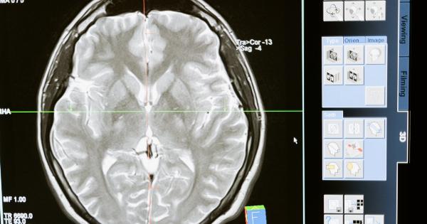
- MRI: Magnetic resonance imaging (MRI) uses magnetic fields to create detailed images of the brain and can help determine the size and location of a hematoma.
- Cerebral Angiography: This procedure involves injecting a dye into the blood vessels of the brain and taking X-ray images to detect any abnormalities or bleeding.
Treatment Options for Syndoclide Brain Hematoma
The appropriate treatment for syndoclide brain hematoma depends on several factors, including the size, location, and severity of the hematoma, as well as the overall health of the patient. Treatment options may include:.
- Surgery: In some cases, a surgical procedure may be necessary to remove the hematoma and repair any damaged blood vessels.
- Medication: Certain medications, such as diuretics or anti-seizure drugs, may be prescribed to manage symptoms or prevent complications.
- Observation and Monitoring: For small hematomas that are not causing significant symptoms, close monitoring and observation may be recommended.
- Rehabilitation: After treatment, rehabilitation therapy may be necessary to help the patient regain lost skills and recover as much functionality as possible.
Complications and Prognosis
Syndoclide brain hematoma can lead to various complications, including:.
- Brain damage: The increased pressure from the hematoma can cause damage to brain tissue, leading to cognitive impairments or physical disabilities.
- Stroke: A brain hematoma can disrupt blood flow to certain areas of the brain, increasing the risk of a stroke.
- Seizures: Hematomas can irritate the brain and trigger seizures.
- Coma: In severe cases, syndoclide brain hematoma can result in a coma or persistent vegetative state.
The prognosis for syndoclide brain hematoma depends on several factors, including the size and location of the hematoma, the patient’s overall health, and the timeliness of treatment.
Prompt medical intervention and appropriate treatment can greatly improve the chances of a favorable outcome.
Prevention of Syndoclide Brain Hematoma
While it may not be possible to prevent all cases of syndoclide brain hematoma, there are certain measures individuals can take to reduce the risk:.
- Wear protective headgear during activities that carry a higher risk of head injury, such as sports or job-related tasks.
- Manage blood pressure levels through a healthy lifestyle, including regular exercise, a balanced diet, and stress reduction techniques.
- Seek medical attention for any significant head injury, even if symptoms are not immediately apparent.
- Follow appropriate safety precautions when using machinery or participating in activities with a risk of head injury.
Conclusion
Syndoclide brain hematoma is a serious condition that requires timely medical intervention. Understanding the causes, symptoms, diagnosis, and treatment options can help individuals recognize the signs of this condition and seek appropriate care.
Although the prognosis for syndoclide brain hematoma can vary based on several factors, prompt treatment greatly improves the chance of a positive outcome. By taking preventative measures and maintaining a healthy lifestyle, individuals can reduce their risk of developing this potentially life-threatening condition.


