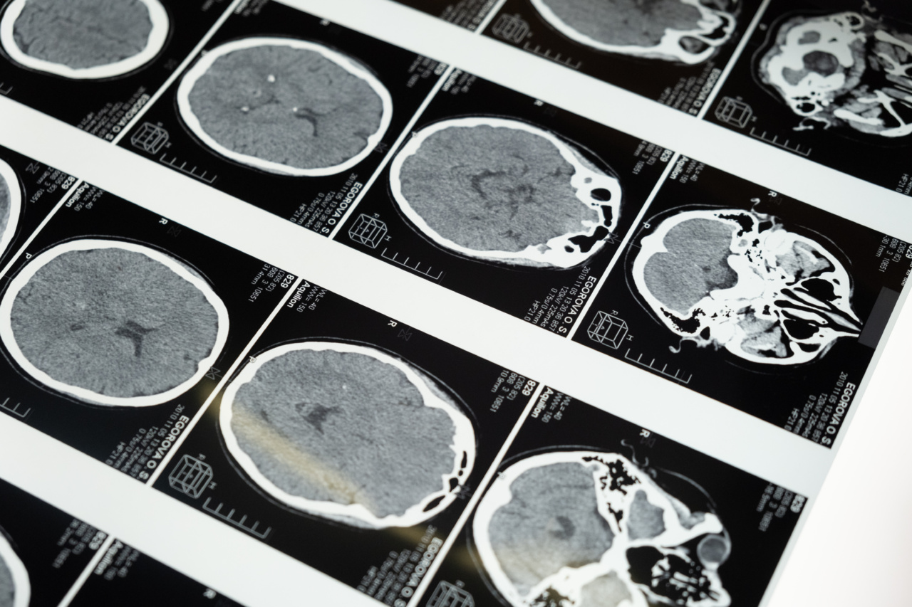
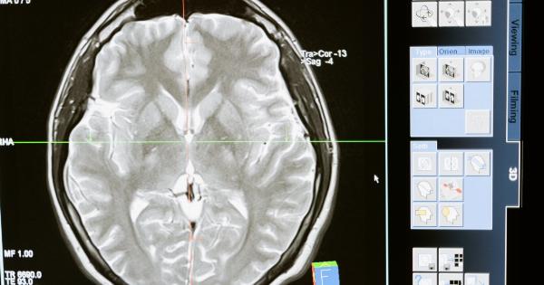
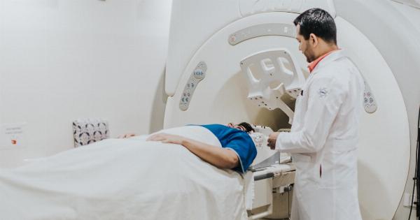
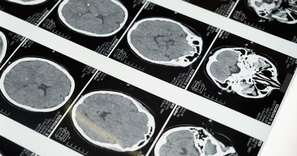
Magnetic Resonance Imaging (MRI) has become an indispensable tool in neuroscience research that has revolutionized our understanding of the underlying brain mechanisms of human behavior.
The study of sex in the brain, also referred to as Neurosexism, has gained tremendous popularity in recent years, primarily due to the growing awareness about gender equity and the impact of sex differences on mental health. MRI provides a non-invasive, safe, and precise method to investigate the structural and functional differences of the brain between males and females.
This article aims to explore the various applications of MRI in studying the sex in the brain and highlights the significant findings in this area of research.
Structural Differences
One of the essential areas of research in neurosexism is the identification of structural differences in the brains of males and females.
MRI allows researchers in this field to probe the brain’s anatomy in vivo, without causing any harm or discomfort to the participant. Several studies have reported that females have a larger hippocampus volume than males, a brain region that plays a vital role in memory and learning.
On the other hand, males have a larger amygdala volume than females, a region that is involved in regulating emotions.
Furthermore, a recent study using Diffusion Tensor Imaging (DTI), another type of MRI technique, showed that the structural connectivity between brain regions was higher in males than females, suggesting that males have a more efficient brain network than females. These findings have significant implications for understanding sex-based differences in brain disorders such as Alzheimer’s Disease and post-traumatic stress disorder (PTSD).
Functional Differences
Besides structural differences, MRI has also been used to identify functional differences in the brains of males and females.
Functional Magnetic Resonance Imaging (fMRI) is a technique that measures the changes in blood flow in the brain when a person performs a specific task, providing insights into which regions of the brain are activated during that task. A study by Cahill et al.
(2001) showed that females have a higher activation in the amygdala and hippocampus when recalling emotionally arousing events, whereas males had a higher activation in the dorsal prefrontal cortex (DPFC), the ventral MPFC, and the dorsal ACC, regions that are involved in cognitive control and emotion regulation. These findings suggest that females have a stronger emotional memory, whereas males have a more cognitive approach towards memory recall.
Another area of research that has gained attention recently is the study of sexual orientation based on brain differences.
A study by Savic and Lindström (2008) showed that the brains of lesbian women respond differently to pheromones than heterosexual women. The fMRI scans showed that the hypothalamus, a region of the brain involved in sexual behavior, was more active in lesbian women when exposed to female-specific pheromones.
Similarly, a study by LeVay (1991) showed that the brain’s anterior hypothalamus was significantly smaller in homosexual men than heterosexual men, suggesting a possible link between sexual orientation and brain anatomy.
Impact of Hormones
Hormones are another critical factor that influences brain development and function. Studies have shown that hormones such as testosterone and estrogen have a significant impact on the structural and functional organization of the brain and behavior.
MRI has been used to investigate the effects of hormones on the brain across the lifespan. For example, a study by Peper et al. (2013) showed that testosterone levels in males were positively correlated with gray matter volume in regions involved in social cognition, such as the MPFC and STS.
On the other hand, studies in females have reported that estrogen levels are associated with increased gray matter volume in regions involved in memory and emotion, such as the hippocampus and amygdala.
Limitations of MRI in Studying Sex in the Brain
Like any other scientific technique, MRI has its limitations when studying sex in the brain.
One of the significant limitations is the reliance on self-report data regarding gender identity and sexual orientation, which can be subjective and prone to biases. Furthermore, the sample size of studies conducted in this area is relatively small, limiting the generalizability of findings.
Finally, it is crucial to acknowledge that structural and functional differences in the brain do not necessarily imply differences in behavior or abilities. Social and cultural factors also play a significant role in shaping gender differences in various cognitive domains.
Applications in Mental Health
The study of sex in the brain using MRI has several applications in mental health.
For example, understanding the structural and functional differences in the brains of males and females can assist in the diagnosis and treatment of various psychiatric disorders, such as depression, anxiety, and post-traumatic stress disorder (PTSD). Furthermore, the use of MRI in studying the effects of hormones on the brain can help in the development of hormone-based therapies for disorders such as polycystic ovary syndrome (PCOS) and transgender healthcare.
Future Directions
MRI has revolutionized our understanding of the brain and sex differences. However, there is still much to learn in this area of research.
Future directions should focus on a larger sample size, including individuals from diverse backgrounds to address potential biases in current research. Furthermore, there is a need for longitudinal studies to investigate changes in brain structure and function across the lifespan and their association with hormones and behavior.
Finally, given the significant role of culture and social factors in shaping gender-related behavior and cognition, future studies should incorporate multidisciplinary approaches to understanding sex in the brain.
Conclusion
MRI provides a powerful tool that has transformed our understanding of the brain and sex differences.
Structural and functional differences in the brain between males and females have been identified, providing insights into various cognitive domains such as memory, emotion, and sexual orientation. Hormones have a significant impact on the structure and function of the brain, and MRI has assisted in understanding this relationship across the lifespan.
Future applications of MRI in this area of research have the potential to assist in the diagnosis and treatment of mental health disorders and the development of hormone-based therapies.
