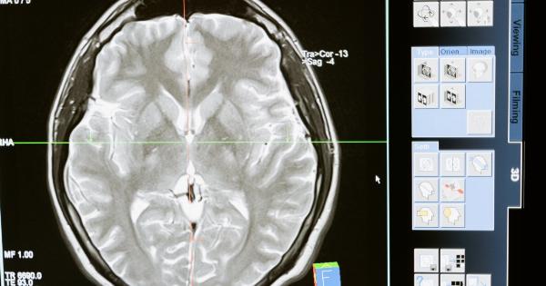
Alzheimer’s disease is a devastating neurodegenerative disease that affects millions of people worldwide. It is characterized by progressive memory loss, cognitive decline, and changes in behavior.
Currently, there is no cure for Alzheimer’s disease, but early detection can significantly improve the management of the condition. Recent research suggests that the eyes may offer valuable insights into Alzheimer’s disease risk through various ocular changes and retinal imaging techniques.
Retinal Imaging and Alzheimer’s Disease
Retinal imaging, a non-invasive technique that captures high-resolution images of the retina, has shown promise in detecting early signs of Alzheimer’s disease.
The retina, which is an extension of the brain, shares many structural and functional similarities with the brain’s neural tissues. Therefore, changes in the retina may reflect the underlying pathological processes occurring in the brain.
One of the key indicators of Alzheimer’s disease is the presence of beta-amyloid plaques, which are abnormal protein deposits in the brain.
Studies have found that these plaques can also be detected in the retina of individuals with Alzheimer’s disease using specialized imaging techniques. By examining the retina, researchers can potentially identify beta-amyloid deposits before cognitive symptoms appear, allowing for early intervention and treatment.
Ocular Changes in Alzheimer’s Disease
Aside from retinal imaging, studies have identified various ocular changes associated with Alzheimer’s disease. These changes include alterations in retinal thickness, changes in retinal blood vessels, and abnormalities in the optic nerve.
These ocular changes may serve as biomarkers for the early diagnosis and monitoring of Alzheimer’s disease.
Researchers have discovered that individuals with Alzheimer’s disease often exhibit thinning of the retinal nerve fiber layer and changes in the macula, which is responsible for central vision.
These changes in retinal thickness can be detected through optical coherence tomography (OCT), a non-invasive imaging technique that provides cross-sectional images of the retina. Monitoring these retinal thickness changes over time may help identify disease progression.
In addition to retinal thickness, alterations in retinal blood vessels have been observed in individuals with Alzheimer’s disease.
Studies have shown that retinal blood vessels have distinct patterns in individuals with Alzheimer’s disease compared to healthy individuals. Analyzing these patterns can potentially provide early indications of the disease and aid in monitoring its progression.
Furthermore, abnormalities in the optic nerve, which connects the retina to the brain, have been identified in individuals with Alzheimer’s disease.
Changes in the optic nerve can be visualized through imaging techniques such as optic coherence tomography and scanning laser polarimetry. These changes may serve as additional biomarkers for tracking Alzheimer’s disease progression and response to treatment.
The Potential of Using Eye Health as a Biomarker
The ability to detect Alzheimer’s disease in its early stages is crucial for developing effective treatments and interventions.
Currently, the diagnosis of Alzheimer’s disease relies on cognitive tests and brain imaging, which may not be sensitive enough for early detection. However, using the eyes as a window into the brain offers the potential for non-invasive, early detection of the disease.
The advantage of using eye health as a biomarker for Alzheimer’s disease is its accessibility and cost-effectiveness. Retinal imaging techniques, such as OCT and specialized camera systems, are readily available in many clinical settings.
These techniques can be easily incorporated into routine eye exams, allowing for widespread screening of individuals at risk for Alzheimer’s disease.
Furthermore, monitoring ocular changes over time may provide valuable insights into disease progression and response to treatment.
By analyzing retinal thickness, retinal blood vessels, and optic nerve abnormalities, clinicians can track the effectiveness of interventions and evaluate the impact of potential Alzheimer’s disease therapies.
Challenges and Future Directions
While the potential of using eye health as a biomarker for Alzheimer’s disease is promising, there are still several challenges that need to be addressed.
First, standardization of imaging protocols and interpretation criteria is essential to ensure consistency and comparability across different studies and clinical settings.
Additionally, more research is needed to establish the specificity and sensitivity of retinal imaging and ocular changes in Alzheimer’s disease.
It is important to determine how well ocular biomarkers can differentiate Alzheimer’s disease from other neurodegenerative diseases or age-related changes in the eye.
Another challenge is developing algorithms and artificial intelligence tools to analyze and interpret the vast amount of data generated from retinal imaging.
These tools can assist clinicians in identifying subtle changes in the retina and provide quantitative measurements for better monitoring and diagnosing Alzheimer’s disease.
As research on the relationship between eye health and Alzheimer’s disease continues to advance, the potential for using the eyes as a biomarker for early detection and understanding the progression of the disease becomes more evident.
The eyes offer a non-invasive and accessible window into the brain, allowing for earlier interventions and improved management of Alzheimer’s disease.


