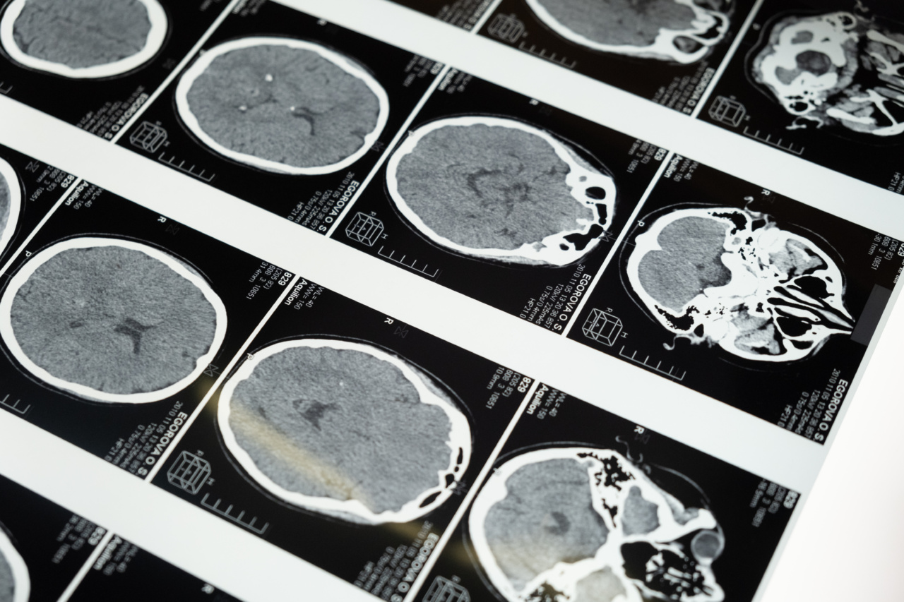
Magnetic Resonance Imaging (MRI) is a medical imaging technique that uses strong magnetic fields and radio waves to generate detailed images of the internal structures of the body.
Unlike other imaging techniques such as X-rays or CT scans, MRI does not use ionizing radiation, making it a safer option for patients. The images produced by MRI can help healthcare professionals diagnose and monitor a wide range of medical conditions.
Principles of Magnetic Resonance Imaging
The underlying principle of MRI involves the behavior of certain atomic nuclei in the presence of a strong magnetic field.
In simple terms, MRI exploits the natural magnetic properties of the body’s hydrogen atoms, which are abundant in water and fat molecules. These atoms have a property called “spin,” which can be visualized as tiny magnets aligning with a strong external magnetic field.
Magnetic Fields and Resonance
The first step in the MRI process involves the patient being placed inside a specially designed cylindrical scanner. This scanner contains powerful magnets that generate a strong and uniform magnetic field.
This strong magnetic field aligns the hydrogen atom spins within the body.
Once the atoms are aligned, a second radiofrequency magnetic field is applied to the body. This radiofrequency field is specifically tuned to a frequency that matches the resonance frequency of the hydrogen atoms.
When the resonance frequency is matched, the atoms absorb energy from the radio waves and become temporarily excited.
Relaxation Process
After the radiofrequency pulse is turned off, the excited hydrogen atoms release the absorbed energy. This energy release is detected by the MRI scanner, and it provides detailed information about the tissues being imaged.
The relaxation process of the excited hydrogen atoms occurs in two stages – the first stage is called “T1 relaxation” and the second stage is called “T2 relaxation.”.
T1 Relaxation
In the T1 relaxation stage, the excited hydrogen atoms release energy and return to their original alignment with the strong external magnetic field. This realignment process is gradual and can take several seconds.
The rate at which the atoms return to their original alignment provides information about the molecular composition and physical properties of the imaged tissues.
T2 Relaxation
In the T2 relaxation stage, the excited hydrogen atoms lose their coherence and their spins begin to interact with each other. The interactions cause the atoms to lose their energy, eventually returning to their original state.
The T2 relaxation process is faster than the T1 relaxation process and is also influenced by the surrounding molecular environment.
Image Formation
To create an image, the MRI scanner manipulates the T1 and T2 relaxation processes. It does this by applying sequences of radiofrequency pulses and magnetic field gradients.
These pulses and gradients result in the emission of weak radio signals from the excited hydrogen atoms, which are then detected by the scanner.
The detected signals are analyzed by a computer, which reconstructs them into detailed images of the imaged tissues.
Different tissues within the body exhibit different signal intensities based on their inherent T1 and T2 relaxation characteristics, allowing for excellent tissue contrast in the resulting images.
Types of MRI Scanners
There are several types of MRI scanners available, each with its own strengths and applications.
The most common type is the “closed” or “tunnel” MRI scanner, where the patient lies on a table that slides into a tunnel-like structure containing the magnet. Closed MRI scanners can sometimes cause discomfort or claustrophobia in patients due to the tight space inside the tunnel.
Another type is the “open” MRI scanner, which is designed to be more comfortable for patients who may experience anxiety or claustrophobia.
Related Article
The science behind magnetic resonance imaging

Open MRI scanners have a larger opening and are suitable for patients who cannot undergo imaging in a closed scanner.
Applications of Magnetic Resonance Imaging
MRI is a versatile imaging technique used in various medical specialties. It provides detailed images of the brain, spinal cord, joints, muscles, and other soft tissues. Some common applications of MRI include:.
1. Neuroimaging: MRI can detect and monitor brain tumors, strokes, neurodegenerative diseases, and other neurological conditions.
2. Orthopedics: MRI is often used to evaluate injuries or disorders of the joints, bones, and muscles. It is particularly useful for diagnosing ligament tears, cartilage damage, and herniated discs.
3. Cardiology: MRI can visualize heart structures and assess heart function, helping in the diagnosis of heart diseases and planning cardiac interventions.
4. Abdominal Imaging: MRI can provide detailed images of the liver, kidneys, pancreas, and other abdominal organs. It is useful in diagnosing tumors, evaluating blood flow, and assessing organ function.
5. Breast Imaging: MRI is sometimes used as a supplemental screening tool for breast cancer, especially in high-risk individuals.
Advantages of Magnetic Resonance Imaging
MRI offers several advantages over other imaging techniques:.
– Non-invasive: MRI does not involve any incisions or injections, making it a non-invasive imaging option.
– Radiation-free: Unlike X-rays and CT scans, which use ionizing radiation, MRI uses magnetic fields and radio waves, eliminating the potential risks associated with radiation exposure.
– Excellent soft tissue contrast: MRI provides detailed images of soft tissues, allowing for the detection and characterization of abnormalities that may not be visible on other imaging modalities.
– Multiplanar imaging: MRI can capture images in multiple planes (e.g., sagittal, coronal, and axial), providing comprehensive views of the imaged structures.
– Functional imaging: Advanced MRI techniques can assess brain function, blood flow, and tissue perfusion, contributing to the understanding of various diseases and conditions.
Limitations and Considerations
While MRI is a highly valuable imaging tool, it also has certain limitations and considerations:.
– Cost: MRI scanners are expensive to install and maintain, which can limit their widespread availability.
– Time-consuming: MRI scans typically take longer to perform than other imaging modalities, and patients need to remain still during the scanning process.
– Contraindications: Some patients with certain metallic implants or devices may not be suitable for MRI due to safety concerns. These implants can cause image artifacts or pose potential risks to the patient.
– Claustrophobia: The close confines of closed MRI scanners can cause anxiety or claustrophobia in some patients. Open MRI scanners are an alternative option for individuals who may experience discomfort.
Conclusion
Magnetic Resonance Imaging is a powerful medical imaging technique that utilizes magnetic fields and radio waves to generate detailed images of the body’s internal structures.
It provides valuable diagnostic information across many medical specialties without exposing patients to ionizing radiation. With ongoing advancements, MRI continues to play a significant role in the diagnosis, treatment, and monitoring of various diseases and conditions.


