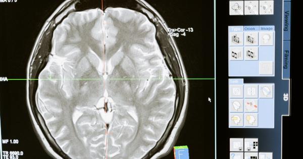
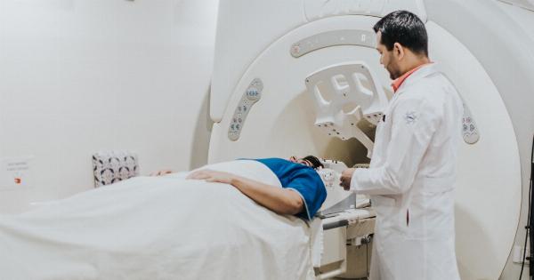
Medical imaging has come a long way, with technological advancements making it easier for healthcare professionals to detect and monitor diseases in patients.
But despite the availability of state-of-the-art equipment and techniques, there are still limitations to what we can see and understand about the human body. Researchers in Japan, however, appear to have made a breakthrough that could change all that.
The Development of Transparent Organs
A team of researchers at the RIKEN Center for Biosystems Dynamics Research in Japan has developed a method to create transparent organs by removing fats and other substances that make up the tissue from specimens.
This technique, known as Clear Lipid-exchanged Acrylamide-hybridized Rigid Imaging/Immunostaining/In situ hybridization-compatible Tissue hYdrogel (CLARITY), allows light to pass through the specimens, making them completely transparent.
While previous attempts at transparency have been made with animal specimens, this is the first time that researchers have been able to create transparent human organs, including the liver, lung, kidney, and pancreas.
This milestone could pave the way for significant advancements in the field of medical imaging, enabling researchers to study organs and tissues in greater detail than ever before.
The Benefits of Transparent Organs
The development of transparent organs has the potential to revolutionize medical imaging, as it provides researchers with a more comprehensive understanding of the human body’s inner workings.
This breakthrough could enable healthcare professionals to better diagnose and treat various diseases and conditions, from cancer and heart disease to Alzheimer’s and Parkinson’s. It could also pave the way for the development of new treatments and therapies, as researchers gain a deeper understanding of the mechanisms behind certain diseases.
Another benefit of transparent organs is that they allow for more accurate and detailed 3D imaging.
This is especially important when studying complex structures such as the brain or organs that are densely packed with cells, as traditional imaging techniques struggle to provide a clear view. With transparent organs, researchers can use advanced imaging methods such as light-sheet microscopy to obtain high-resolution images of the tissues and structures they are studying.
Current Applications of Transparent Organs
While the development of transparent organs is still in its early stages, researchers are already exploring some potential applications of this breakthrough.
One area of focus is cancer research, where transparent organs can help researchers better understand how cancer cells behave and spread throughout the body. With a clearer view of the cancer cells in action, researchers can develop more effective treatment options.
Transparent organs could also lead to more accurate disease modeling.
By studying how diseases develop and progress in transparent tissues, researchers can create more realistic models of disease that can be used to test potential treatments and therapies. This could speed up the drug development process and ultimately lead to better outcomes for patients.
The Future of Medical Imaging
The development of transparent organs is just one example of how medical imaging is advancing.
Researchers are constantly finding new ways to visualize and understand the human body, whether it’s through the use of AI and machine learning or the development of new imaging technologies.
In the years to come, we can expect to see even more breakthroughs in medical imaging, with researchers using these innovative techniques to gain a deeper understanding of the human body and develop new treatments and therapies for a range of diseases and conditions.
Conclusion
The development of transparent organs is a significant breakthrough that could transform the field of medical imaging.
By providing healthcare professionals with a clearer and more comprehensive view of the human body, researchers can better diagnose and treat diseases, ultimately leading to better outcomes for patients. While the technology is still in its early stages, the potential applications of transparent organs are vast, providing hope for the development of new treatments and therapies that could improve the lives of millions of people around the world.


