Doppler ultrasound is a type of ultrasound that uses high-frequency sound waves to measure blood flow in different parts of the body.
In pregnancy, it can be used to monitor the fetal heartbeat and blood flow in the umbilical cord, placenta, and fetal brain. In this guide, we will discuss everything you need to know about Doppler ultrasound in pregnancy.
What is Doppler Ultrasound?
Doppler ultrasound is a type of ultrasound that uses sound waves to measure the velocity and direction of blood flow in the body. It was first introduced in the 1950s and has since become a widely used medical imaging technology.
Doppler ultrasound is non-invasive and does not involve radiation, making it safe for both the mother and the fetus.
How is Doppler Ultrasound Used in Pregnancy?
Doppler ultrasound can be used in pregnancy to monitor the fetal heartbeat and blood flow in the umbilical cord, placenta, and fetal brain.
It can also be used to assess the risk of pre-eclampsia, a serious pregnancy complication that can lead to high blood pressure, organ damage, and other health problems for the mother and baby.
When is Doppler Ultrasound Performed?
Most pregnant women will have at least one ultrasound exam during their pregnancy. The timing and frequency of these exams may vary depending on the health of the mother and the fetus.
Doppler ultrasound is typically performed in the second and third trimesters of pregnancy, although it may be indicated earlier in certain cases, such as in high-risk pregnancies.
What are the Risks of Doppler Ultrasound in Pregnancy?
Doppler ultrasound is considered safe for both the mother and the fetus. It does not involve radiation and is non-invasive.
However, excessive use of Doppler ultrasound may be associated with a slight increase in the risk of low birth weight and preterm birth.
What are the Benefits of Doppler Ultrasound in Pregnancy?
The benefits of Doppler ultrasound in pregnancy include: – Monitoring fetal heartbeat and blood flow – Assessing the risk of pre-eclampsia – Detecting fetal abnormalities – Evaluating fetal growth and development.
Related Article
Understanding Pregnancy Doppler Ultrasound

How is Doppler Ultrasound Performed?
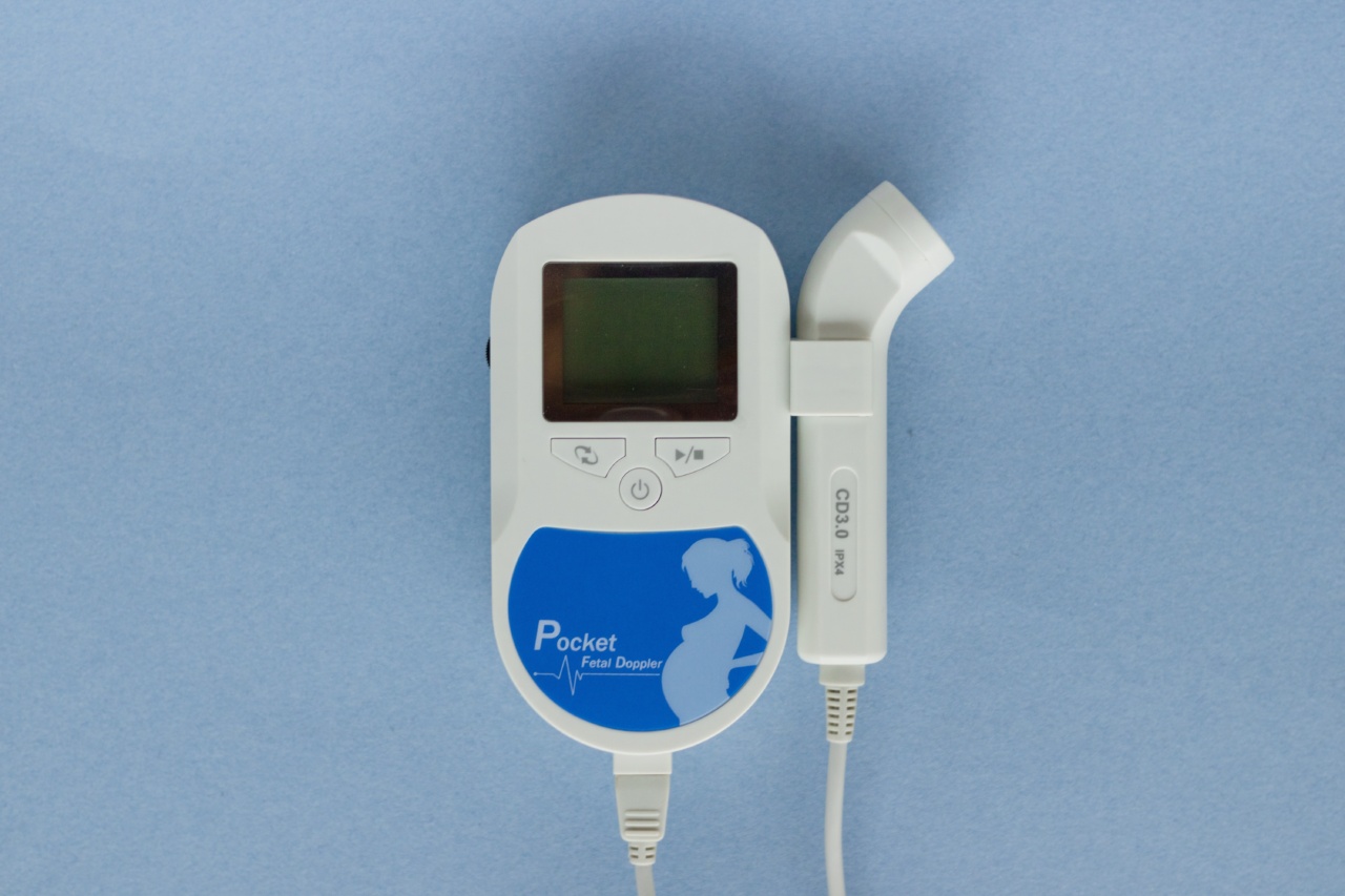
Doppler ultrasound is performed using a handheld device called a transducer, which is placed on the abdomen of the mother. The transducer emits high-frequency sound waves that bounce off the tissues and blood vessels in the body.
The Doppler effect is then used to measure the velocity and direction of blood flow.
What Can Doppler Ultrasound Show?
Doppler ultrasound can show: – Fetal heartbeat – Blood flow in the umbilical cord, placenta, and fetal brain – Signs of pre-eclampsia – Fetal growth and development – Abnormalities or complications in the pregnancy.
What Happens During a Doppler Ultrasound Exam?
During a Doppler ultrasound exam, the mother will lie on a table and a gel will be applied to her abdomen. The transducer will be placed on the gel and moved around to get different views of the fetus and its blood vessels.
The exam usually takes between 30 and 60 minutes.
What Do the Results of a Doppler Ultrasound Exam Mean?
The results of a Doppler ultrasound exam can help healthcare providers: – Assess fetal well-being – Monitor fetal growth and development – Detect fetal abnormalities or complications – Determine the risk of pre-eclampsia.
What Happens if a Problem is Detected During a Doppler Ultrasound Exam?
If a problem is detected during a Doppler ultrasound exam, further tests and monitoring may be recommended.
These may include: – Additional ultrasound exams – Fetal echocardiography – Non-stress test – Biophysical profile – Amniocentesis.
Conclusion
Doppler ultrasound is a safe and non-invasive way to monitor fetal well-being and assess the risk of pre-eclampsia in pregnancy. Healthcare providers may recommend one or more Doppler ultrasound exams depending on the health of the mother and the fetus.
If a problem is detected during a Doppler ultrasound exam, further tests and monitoring may be recommended to ensure the best possible outcomes for both the mother and the baby.


