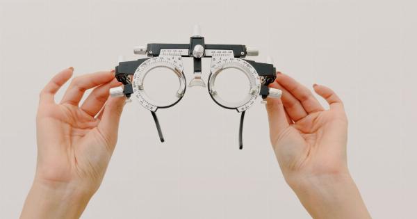
Have you been experiencing difficulty seeing things or noticing changes in your eyesight? It could be time to discover if you have any visual impairments.
Visual impairments can vary in severity, and early detection can lead to prompt treatment and management.
In this article, we will look at various tests that can help you discover any visual impairments you may have. Let’s get started!.
Snellen Eye Chart Test
The Snellen Eye Chart Test is a widely used test to assess a person’s visual acuity. It is a chart with letters that decrease in size as you move down the chart.
The viewer stands at a distance of twenty feet from the chart and is asked to read the smallest line of letters possible.
The results of the test determine the clarity of your vision and if you need corrective lenses.
A score of 20/20 is considered excellent vision, but if you have a score below this, it means you have a visual impairment that requires correction.
Eye Dilation Test
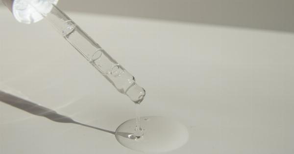
An eye dilation test involves dilating the pupils with special eye drops to evaluate the back portion of the eye. Ophthalmologists use this test to assess the optic nerves, blood vessels, and retina of an individual.
The test helps detect several eye diseases and conditions such as cataracts, macular degeneration, and glaucoma.
The test is painless and takes about 20-30 minutes, but the dilation effect could last a few hours, making tasks like driving or reading difficult for a while.
Visual Field Test
A visual field test assesses the full horizontal and vertical range of vision.
The test helps to identify any regions of vision loss in both eyes due to glaucoma, tumor, optic nerve damage, or other eye conditions.
In a visual field test, the examiner uses a machine to show light patterns at various positions in the viewer’s visual field. The viewer is asked to respond by pressing a button or indicating the presence of light. The results of this test help evaluate the extent and severity of any visual field loss.
Retinal Examination Test
The retinal examination test is a quick and thorough evaluation of the retina through the use of special equipment. The test helps identify any retinal abnormalities or conditions that could cause visual impairment.
Conditions like age-related macular degeneration (AMD), diabetic retinopathy, and retinal detachment can be detected by the retinal examination test.
The test involves shining a bright light in the eyes, and the examiner views the retina through a specialized lens. The test can be done at an eye clinic or doctor’s office.
Color Blindness Test
A color blindness test is done to evaluate whether a person has difficulty distinguishing certain colors. The test assesses the production and perception of color.
The Ishihara test, a widely used color blindness test, uses a series of plates with colored dots to determine if an individual can recognize numbers hidden in the dots.
The test is quick, straightforward, and painless. It can be administered at home or at a clinic, and the results can identify color blindness or color vision deficiencies in individuals.
Near Vision Acuity Test
A near vision acuity test evaluates a person’s ability to read near objects. The test helps identify any hyperopia or presbyopia that occurs when the eye lens loses flexibility and results in farsightedness.
The test involves reading a standard text with small print at a specific distance.
Corrective lenses can be prescribed if the test results identify any near vision impairment.
Slit lamp Examination Test
A slit lamp examination test is a specialized test used to evaluate the structures of the eye. It uses a microscope with a narrow beam of light that shines on the eye.
The examiner views the eye structures, such as the cornea, iris, lens, and sclera, for abnormalities such as clouding, scratches, glaucoma, and cataracts.
The test is non-invasive and takes about 15-20 minutes to complete.
Optical Coherence Tomography Test
The Optical coherence tomography (OCT) test uses light waves to create high-resolution images of the retina.
The test helps identify retinal diseases such as macular degeneration, diabetic retinopathy, and glaucoma.
The test is similar to an ultrasound test and uses light waves instead of sound waves. The OCT test takes about 10 to 15 minutes to complete, and results provide detailed information about the health and thickness of the retina.
Retinal Photography Test
Retinal photography involves using a specialized camera to take digital images of the retina. The test helps identify any abnormalities, inflammations, or eye diseases that may require treatment.
The test results can help track the progress of treatments planned.
Retinal photography is non-invasive and takes less than 10 minutes to complete. The images captured in the test are high-resolution and provide clear details of the retina.
Visual Acuity Test for Children
Visual Acuity Tests for children are conducted to assess children’s vision and to identify any visual impairments that may need correction.
Many children with vision impairments may not be aware of their difficulties and struggle with reading, learning, and engaging in their day to day activities.
Visual Acuity tests for children are similar to the Snellen test but use age-appropriate letters and characters. Corrective lenses can be prescribed if the test identifies any visual impairments.


