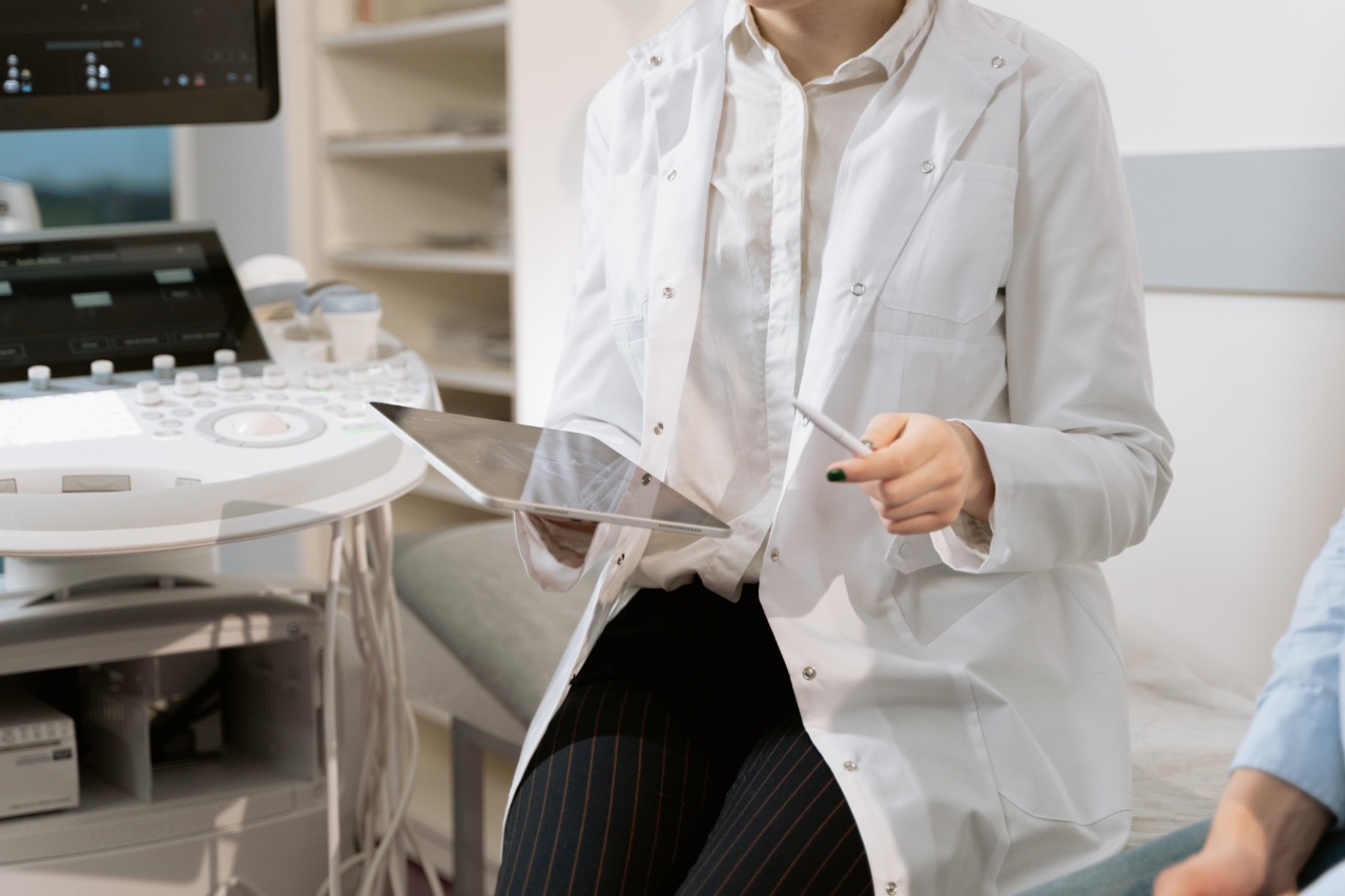
Breast cancer is one of the leading causes of death among women worldwide. Early detection is key to the successful treatment of breast cancer. One of the most effective tools used for detecting breast cancer is ultrasound.
In recent years, there have been groundbreaking technologies in breast ultrasound that have made the detection and diagnosis of breast cancer more accurate and efficient. These technologies include contrast-enhanced ultrasound, automated breast ultrasound, and elastography.
Contrast-Enhanced Ultrasound (CEUS)
CEUS is a technology that involves the use of ultrasound contrast agents that are injected into the patient’s bloodstream. These contrast agents help to enhance the images produced during ultrasound scans.
The use of CEUS has been shown to improve the detection and characterization of breast lesions.
CEUS has been found to be particularly useful in the assessment of breast lesions that are difficult to visualize using conventional ultrasound.
These include small lesions, lesions in dense breast tissue, and lesions that are located close to the chest wall.
CEUS is a safe and non-invasive procedure that can be performed in an outpatient setting. It does not involve the use of ionizing radiation or contrast agents that contain iodine.
This makes it an ideal imaging modality for patients who are allergic to iodine or who have renal insufficiency.
Automated Breast Ultrasound (ABUS)
ABUS is a technology that involves the use of a specialized ultrasound system to perform a 3D ultrasound scan of the breast. The images obtained using ABUS are then analyzed using computer algorithms to detect and characterize breast lesions.
ABUS is particularly useful in the screening of women with dense breast tissue. Dense breast tissue can make it difficult to detect breast lesions using conventional mammography.
ABUS has been shown to be more sensitive than mammography in the detection of breast cancer in women with dense breast tissue.
ABUS is a non-invasive procedure that does not involve the use of ionizing radiation. It is also less uncomfortable for patients than mammography, as it does not involve breast compression.
Elastography
Elastography is a technology that involves the use of ultrasound to assess the stiffness of breast tissue. Breast cancer is typically associated with increased tissue stiffness.
Elastography can help to identify areas of increased tissue stiffness, which may indicate the presence of breast cancer.
Elastography is particularly useful in the assessment of breast lesions that are difficult to characterize using conventional ultrasound. These include lesions that are small or that have irregular shapes or margins.
Elastography is a safe and non-invasive procedure that does not involve the use of ionizing radiation or contrast agents.
Shear Wave Elastography
Shear Wave Elastography (SWE) is a technology that is used to assess the stiffness of breast tissue. It uses focused sound waves to produce elastic waves in breast tissue. These waves are then measured to determine the stiffness of the tissue.
SWE is particularly useful in the assessment of breast lesions that are difficult to characterize using conventional ultrasound. It has been shown to be more accurate than elastography in the differentiation of benign and malignant breast lesions.
SWE is a safe and non-invasive procedure that does not involve the use of ionizing radiation or contrast agents.
Automated Breast Volume Scanner (ABVS)
The Automated Breast Volume Scanner (ABVS) is a technology that is used to create a 3D image of the breast. It uses a robotic arm to move the ultrasound probe around the breast and create a series of images.
These images are then reconstructed into a 3D image of the breast.
ABVS is particularly useful in the assessment of breast lesions in women with dense breast tissue. It has been shown to be more accurate than mammography in the detection of breast cancer in women with dense breast tissue.
ABVS is a non-invasive procedure that does not involve the use of ionizing radiation. It is also less uncomfortable for patients than mammography, as it does not involve breast compression.
Elastography-Enhanced Ultrasound (EEUS)
Elastography-Enhanced Ultrasound (EEUS) is a technology that combines elastography and CEUS to improve the detection and characterization of breast lesions.
It involves the injection of an ultrasound contrast agent into the patient’s bloodstream, followed by an assessment of tissue stiffness using elastography.
EEUS is particularly useful in the detection and diagnosis of small breast lesions. It has been shown to be more accurate than conventional ultrasound in the detection of breast cancer in women with dense breast tissue.
EEUS is a safe and non-invasive procedure that does not involve the use of ionizing radiation or contrast agents that contain iodine.
Conclusion
The use of ultrasound in the detection and diagnosis of breast cancer has been revolutionized by the development of these groundbreaking technologies.
CEUS, ABUS, elastography, shear wave elastography, ABVS, and EEUS have all been shown to improve the accuracy and efficiency of breast cancer detection and diagnosis. These technologies are safe, non-invasive, and offer an alternative imaging modality to mammography. The use of these technologies has the potential to save countless lives by allowing for earlier detection and treatment of breast cancer.


