The female reproductive system is an amazing and complex network of organs that work together to create life. However, this intricate system can be susceptible to a range of pelvic disorders that can cause pain, discomfort, and even infertility.
Understanding the anatomy of the female pelvic region is important in the diagnosis and treatment of such disorders.
What is the Female Pelvic Region?
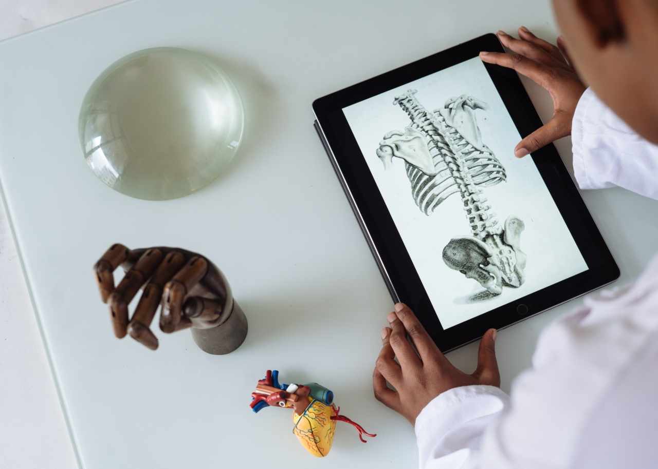
The female pelvis is the bony structure that surrounds the lower part of the abdomen, connecting the spine and the hips. It consists of several individual bones that are joined together with strong ligaments.
The female pelvic region contains the reproductive organs, including the ovaries, uterus, and fallopian tubes, as well as the bladder, rectum, and various muscles and blood vessels.
Anatomy of the Female Reproductive System
The female reproductive system is made up of several organs that work together to produce, transport, and nourish eggs, as well as create a suitable environment for a growing fetus. These organs include:.
Ovaries
The ovaries are two small, almond-shaped organs that are located on each side of the uterus. They are responsible for producing and releasing eggs during ovulation and are also the primary source of hormones such as estrogen and progesterone.
Uterus
The uterus is a pear-shaped organ that is located in the pelvis between the bladder and rectum.
It is the site of fetal development during pregnancy and is composed of three layers: the innermost layer called the endometrium, the middle layer made up of smooth muscle tissue, and the outer layer known as the serosa.
Fallopian Tubes
The fallopian tubes are two thin tubes that extend from each side of the uterus toward the ovaries. They provide a pathway for the eggs to travel from the ovaries to the uterus and are also the site of fertilization between the egg and sperm.
Vagina
The vagina is a muscular tube that connects the uterus to the external genitalia. It serves as a passageway for menstrual blood and is the site of sexual intercourse and childbirth.
Anatomy of the Pelvic Floor
The pelvic floor refers to the group of muscles, ligaments, and connective tissue that support the pelvic organs and control bladder and bowel function. The pelvic floor is made up of three layers:.
Deep Layer
The deep layer includes the muscles that surround the openings of the urethra, vagina, and rectum. These muscles provide support to the pelvic organs and control bladder and bowel function.
Middle Layer
The middle layer consists of muscles that attach to the pubic bone and the tailbone. These muscles work together to support the pelvic organs and help maintain continence.
Superficial Layer
The superficial layer includes the muscles that cover the pelvic bones and support the organs from below. These muscles are involved in sexual function and play a role in helping to maintain continence.
Pelvic Disorders in Women
There are several common pelvic disorders that affect women. These include:.
Endometriosis
Endometriosis is a condition in which the tissue that normally lines the inside of the uterus grows outside of it. This can cause pain, heavy periods, and infertility.
Uterine Fibroids
Uterine fibroids are non-cancerous growths that develop in the uterus. They can cause heavy bleeding, pain, and discomfort.
Urinary Incontinence
Urinary incontinence is the involuntary loss of urine. It can be caused by weakness of the pelvic floor muscles, nerve damage, or other factors.
Pelvic Organ Prolapse
Pelvic organ prolapse occurs when the muscles and tissues that support the pelvic organs become weak or damaged, causing the organs to move from their normal position. This can result in discomfort, pressure, and urinary or bowel problems.
Polycystic Ovary Syndrome (PCOS)
PCOS is a hormonal disorder in which the ovaries produce too much androgen. This can cause irregular periods, acne, and infertility.
Understanding the Anatomy is Key to Diagnosis and Treatment
Understanding the anatomy of the female pelvic region is important in the diagnosis and treatment of pelvic disorders. Medical professionals can use imaging tests such as ultrasounds, CT scans, or MRIs to view the pelvic organs better.
In many cases, they may also perform a pelvic exam to assess the health and function of the pelvic organs and the pelvic floor.
Treatment will depend on the specific disorder and its severity. Options may include medication, surgery, physical therapy, or a combination of treatments.
In many cases, pelvic floor exercise can be an effective way to improve muscle strength and support the pelvic organs, improving urinary incontinence and other pelvic health conditions.
It is important for women to understand their anatomy and any potential pelvic disorders they may have. By working closely with healthcare professionals, women can manage these conditions and maintain optimal pelvic health.


