Vision disorders can be caused by various factors such as age, disease, injury, genetics, and environmental factors. They can range from mild to severe, and some of them can be difficult to diagnose without proper testing.
However, diagnostic imaging has made it possible to detect and identify visual disorders more accurately and efficiently.
Magnetic Resonance Imaging (MRI) for Vision Disorders
Magnetic resonance imaging (MRI) is a diagnostic imaging technique that uses a powerful magnet and radio waves to produce detailed images of internal body structures, including the eye and brain.
MRI is particularly useful for identifying various vision disorders such as optic nerve damage, pituitary gland tumors, and traumatic injury to the eye or brain.
In an MRI test for the eye, a special surface coil is placed over the eye and the patient is asked to lie still while the machine takes images. This test can help diagnose optic neuritis, macular degeneration, and other eye conditions.
MRI can also be used to diagnose brain injuries that result in vision problems, such as concussions and strokes.
Computed Tomography (CT) for Vision Disorders
Computed tomography (CT), also known as computerized axial tomography (CAT) scan, is a diagnostic imaging technique that uses X-rays and computer software to produce detailed images of internal body structures.
CT is particularly useful for identifying various vision disorders such as tumors, hemorrhages, and infections in the eye and brain.
In a CT test for the eye, the patient is asked to lie still on a table while the machine takes images. This test can help diagnose tumors in the eye or optic nerve, as well as infections such as orbital cellulitis.
CT is also useful for identifying brain tumors that cause vision problems.
Ultrasound for Vision Disorders
Ultrasound is a diagnostic imaging technique that uses high-frequency sound waves to produce images of internal body structures.
Ultrasound is particularly useful for identifying various vision disorders such as retinal detachment, cataracts, and glaucoma.
In an ultrasound test for the eye, a special probe is placed on the eye while the patient looks in different directions. This test can help diagnose abnormalities in the eye such as retinal detachment or cataracts.
Ultrasound is also useful for measuring the thickness of the optic nerve, which can be an indicator of glaucoma.
Ophthalmoscopy for Vision Disorders
Ophthalmoscopy is a diagnostic technique that uses a special instrument called an ophthalmoscope to examine the inside of the eye.
Ophthalmoscopy is particularly useful for identifying various eye disorders such as diabetic retinopathy, hypertensive retinopathy, and macular degeneration.
Related Article
Vision impairments: Diagnostic images for common diseases

In an ophthalmoscopy test, the patient’s pupils are dilated with eye drops and the ophthalmologist examines the inside of the eye using an ophthalmoscope.
This test can help diagnose various eye disorders, including problems with the retina, optic nerve, and blood vessels in the eye.
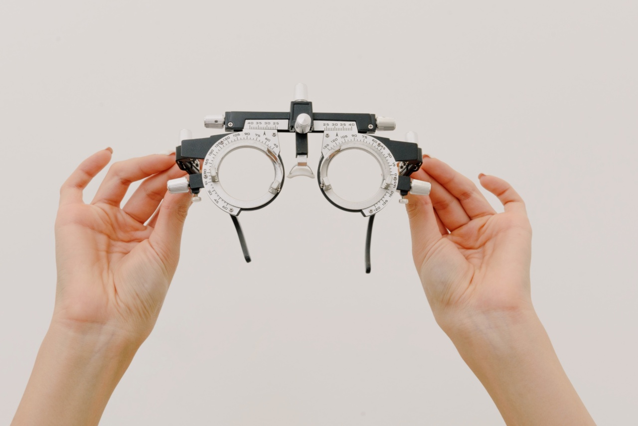
Visual Acuity Tests for Vision Disorders
Visual acuity tests are diagnostic tests that measure the clarity or sharpness of a person’s vision. They are commonly used to screen for vision disorders such as myopia, hyperopia, and astigmatism.
In a visual acuity test, the patient is asked to read letters or numbers from a chart located at a specific distance. This test can help diagnose various vision disorders and determine if corrective lenses are needed for the patient.
Color Vision Tests for Vision Disorders
Color vision tests are diagnostic tests that measure a person’s ability to distinguish between colors. They are commonly used to screen for color vision disorders such as color blindness.
In a color vision test, the patient is shown a series of colored plates and asked to identify the numbers or shapes within the plates. This test can help diagnose various color vision disorders and determine the severity of the condition.
Electroretinography (ERG) for Vision Disorders
Electroretinography (ERG) is a diagnostic test that measures the electrical response of the retina to light. ERG is particularly useful for identifying various vision disorders such as retinitis pigmentosa and age-related macular degeneration.
In an ERG test, the patient’s pupil is dilated with eye drops and the patient is asked to look at a flashing light. A special electrode is placed on the surface of the eye and a computer records the electrical responses of the retina.
This test can help diagnose various retinal disorders and determine the severity of the condition.
Visual Field Testing for Vision Disorders
Visual field testing is a diagnostic test that measures the range of a person’s peripheral or side vision. It is commonly used to screen for vision disorders such as glaucoma and brain tumors.
In a visual field test, the patient is asked to look straight ahead while small flashes of light are presented at different locations around the edge of their vision.
The patient signals when they see the flashes, and the results are recorded on a chart. This test can help diagnose various vision disorders and determine the severity of the condition.
Slit-Lamp Examination for Vision Disorders
Slit-lamp examination is a diagnostic technique that uses a special instrument called a slit-lamp to examine the front and back of the eye.
Slit-lamp examination is particularly useful for identifying various eye disorders such as cataracts and corneal ulcers.
In a slit-lamp examination, the patient’s pupils are dilated with eye drops and the patient sits in front of the microscope-like instrument. The ophthalmologist examines the eye using a beam of light from the slit-lamp and a special lens.
This test can help diagnose various eye disorders and determine the severity of the condition.


