Varicose veins are a prevalent medical condition that affects around 23 percent of adults. This condition causes enlarged, twisted veins, which appear blue or purple.
Symptoms of varicose veins include aching, swelling, and heaviness in the legs, and can cause irreversible damage if not treated promptly. To diagnose this condition, doctors often rely on ultrasound imaging. In this article, we will discuss how ultrasound imaging helps in understanding varicose veins.
What are varicose veins?
Varicose veins occur when blood pools in the veins instead of flowing upwards towards the heart. This occurs due to weakened or damaged vein walls which prevent proper blood flow.
Varicose veins often appear on the legs, but they can develop in other parts of the body as well. Risk factors associated with varicose veins include age, gender, obesity, pregnancy, and a family history of the condition.
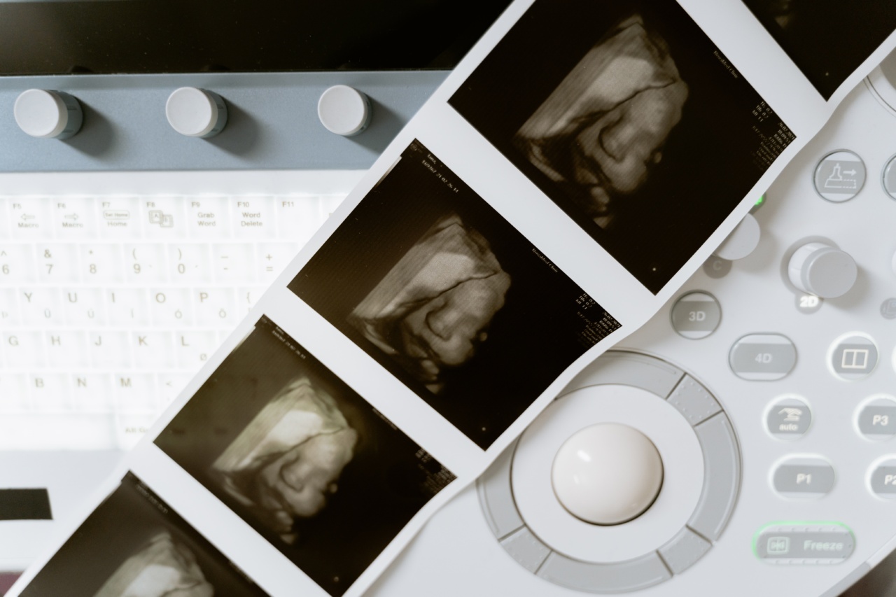
How does ultrasound imaging work?
Ultrasound imaging uses high-frequency sound waves to produce images of the inside of the body. During an ultrasound procedure, a technician uses a transducer, a small handheld device that emits sound waves, to capture images of the veins.
By analyzing these images, doctors can determine the location and severity of varicose veins.
Types of ultrasound imaging for varicose veins
There are two types of ultrasound imaging used to diagnose varicose veins:.
Related Article
How ultrasound imaging helps diagnose varicose veins

- Doppler ultrasound: This method uses sound waves that bounce off blood cells to produce an image of the blood vessels. It helps to determine the direction and speed of blood flow, which can help diagnose arterial and venous diseases.
- Color duplex ultrasound: This technique uses both sound waves and color imaging to produce a real-time image of blood flow in the body’s veins. It can help to detect abnormalities in the venous system such as blood clots, venous insufficiency, and varicose veins.
What can ultrasound imaging reveal about varicose veins?
Ultrasound imaging provides valuable information on the underlying cause of varicose veins. This method can determine:.
- The location of the varicose veins
- The depth and size of the veins
- The severity of the condition
- The direction of blood flow within the veins
- Any blood clot formation in the veins
- The presence of venous insufficiency
Benefits of ultrasound imaging for varicose veins
Ultrasound imaging is a non-invasive and painless procedure, making it an excellent diagnostic tool for varicose veins. It allows doctors to diagnose the condition accurately and provide appropriate treatment. Benefits of ultrasound imaging include:.
- No radiation exposure
- No need for sedation or anesthesia
- No recovery time needed
- The ability to obtain real-time imaging of the veins
What does a patient expect during the ultrasound procedure?
During the ultrasound procedure, the patient lies on a table, and a technician applies a gel to the skin over the area to be examined. The gel helps the transducer make a secure contact with the skin, which enables clear imaging.
The technician moves the transducer over the skin, and sound waves transmitted from the device produce images that appear on a monitor. After the procedure, the patient can resume normal activities immediately.
Conclusion
Varicose veins are a common condition that requires timely intervention to prevent complications. Ultrasound imaging is a non-invasive and painless method that helps to diagnose varicose veins accurately.
By providing detailed images of the veins’ structure, direction of blood flow, and any blood clot formation, ultrasound imaging helps doctors prescribe appropriate treatment. Given the benefits of this diagnostic method, patients should not hesitate to undergo the procedure to ensure early detection and treatment of varicose veins.


