Prostate cancer is the second most common type of cancer in men worldwide, with over a million new cases reported every year.
Early detection and accurate diagnosis play a crucial role in improving the prognosis and survival rates of prostate cancer patients. Magnetic Resonance Imaging (MRI) has emerged as a powerful non-invasive tool for diagnosing and staging prostate cancer, offering high accuracy compared to traditional methods.
This article explores the advancements in MRI technology and its application in diagnosing prostate cancer with exceptional precision.
Advancements in MRI Technology
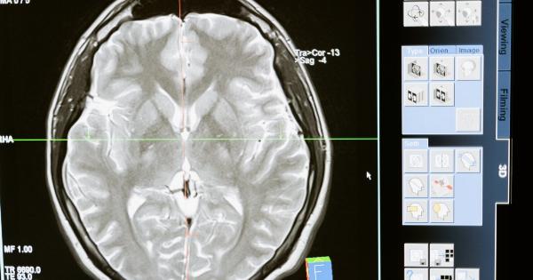
MRI technology has witnessed significant advancements over the years, enabling radiologists to obtain highly detailed images of the prostate gland.
High-resolution MRI scans provide a clearer visualization of the prostate tissue, allowing for better detection and characterization of cancerous lesions. The following are key developments that have led to improved accuracy in diagnosing prostate cancer using MRI:.
1. Multi-Parametric MRI (mpMRI)
Multi-Parametric MRI (mpMRI) combines multiple imaging sequences to provide a comprehensive evaluation of the prostate gland. It includes T2-weighted imaging, diffusion-weighted imaging (DWI), and dynamic contrast-enhanced imaging (DCE), among others.
By assessing different aspects of the tissue, mpMRI significantly enhances the accuracy of prostate cancer diagnosis.
2. Diffusion-Weighted Imaging (DWI)
DWI measures the movement of water molecules in tissue, allowing for the differentiation between cancerous and non-cancerous cells.
Malignant tissue often exhibits restricted diffusion due to the increased cellularity and disrupted cellular structure associated with cancer. DWI has demonstrated high sensitivity and specificity in detecting prostate cancer.
3. Dynamic Contrast-Enhanced Imaging (DCE)
DCE MRI involves the injection of a contrast agent and acquiring sequential images to assess the blood flow and vascularity of the prostate gland.
Cancerous lesions often exhibit increased vascularity due to angiogenesis, and DCE helps in identifying suspicious regions for targeted biopsies. By combining DCE with other MRI sequences, the accuracy of prostate cancer diagnosis can be significantly improved.
4. Prostate Imaging Reporting and Data System (PI-RADS)
The Prostate Imaging Reporting and Data System (PI-RADS) is a standardized scoring system used to evaluate and report prostate MRI findings.
It provides a structured approach for radiologists to assign a score indicating the likelihood of clinically significant prostate cancer. PI-RADS has been widely adopted, enabling better communication and consistency in interpreting MRI results.
High Accuracy Diagnosis
MRI has demonstrated high accuracy in diagnosing prostate cancer, surpassing traditional diagnostic techniques such as transrectal ultrasound-guided biopsy.
A combination of mpMRI sequences, including T2-weighted imaging, DWI, and DCE, enables radiologists to identify suspicious lesions and evaluate their aggressiveness. Here are the key factors contributing to the high accuracy of MRI in prostate cancer diagnosis:.
1. Improved Visualization
High-resolution MRI provides detailed images of the prostate gland, enabling radiologists to visualize cancerous lesions with excellent clarity.
The ability to accurately identify and delineate tumors is crucial for planning treatment and monitoring disease progression.
2. Detection of Small Lesions
MRI has the capability to detect smaller lesions that may be missed by other imaging modalities. With the advent of mpMRI, the sensitivity of detecting clinically significant prostate cancer has significantly increased.
3. Targeted Biopsies
By identifying suspicious regions on MRI, urologists can perform targeted biopsies, leading to increased accuracy and reduced unnecessary biopsies.
MRI-guided biopsies can improve the selection of patients for active surveillance versus immediate treatment.
4. Staging and Localization
MRI is invaluable in accurately staging prostate cancer, helping clinicians determine the extent of the disease and its spread beyond the prostate gland.
It aids in treatment planning and decision-making, ensuring appropriate therapies are administered.
5. Monitoring Treatment Response
Follow-up MRI scans can assess the response to treatment, enabling clinicians to identify any residual or recurrent tumor. This allows for timely intervention and adjustment of treatment plans if necessary.
Challenges and Future Directions
While MRI offers high accuracy in diagnosing prostate cancer, there are challenges that need to be addressed for further improvement:.
1. Standardization
There is a need for standardized protocols and reporting systems to ensure consistency across different centers and radiologists. This facilitates communication and comparison of results, enhancing the overall accuracy of prostate cancer diagnosis.
2. Cost
MRI can be relatively expensive, making widespread implementation challenging in some healthcare settings. Efforts to reduce costs and increase accessibility will play a critical role in improving the diagnosis and management of prostate cancer.
3. Advanced Imaging Techniques
Ongoing research is focused on developing advanced imaging techniques to further improve the accuracy of prostate cancer diagnosis.
This includes the use of artificial intelligence algorithms to aid in lesion detection and characterization, as well as the exploration of novel imaging biomarkers.
Conclusion
MRI has revolutionized the diagnosis of prostate cancer, offering high accuracy and facilitating early detection and precise characterization of tumors.
The advancements in MRI technology, such as mpMRI, DWI, and DCE, have significantly improved its diagnostic capabilities. With continued research and standardization efforts, MRI will continue to play a pivotal role in effectively diagnosing and managing prostate cancer, thereby improving patient outcomes.


