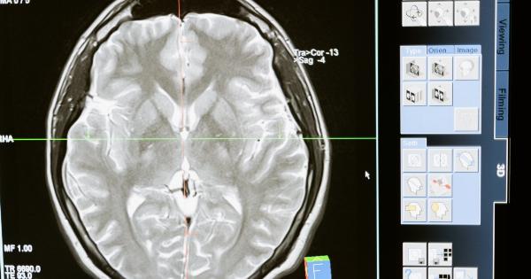
Magnetic resonance imaging (MRI) is a sensitive and non-invasive imaging technique used to diagnose and stage prostate cancer.
Nonetheless, the diagnostic accuracy can be improved using polyparametric MRI (ppMRI), which combines different MRI sequences and additional techniques to provide more precise and comprehensive information. The aim of this article is to describe the advantages and limitations of ppMRI for prostate cancer diagnosis and management.
How Does ppMRI Work?
ppMRI relies on the combination of three or more MRI sequences, which generate different types of images and enhance different properties of the prostate tissue. The core sequences used in ppMRI are:.
T2-Weighted Imaging (T2WI)
T2WI is the standard MRI sequence for prostate imaging and is based on the signal intensity of water molecules in the tissue. It provides anatomical information on the prostate gland, including its size, shape, and zonal anatomy.
T2WI is particularly useful for detecting prostate cancer, as cancerous tissues usually have a lower signal intensity than normal tissues.
Diffusion-Weighted Imaging (DWI)
DWI is a technique that measures the random motion of water molecules within the tissue. Cancerous tissues usually have restricted diffusion due to the increased cell density, and appear as high signal intensity regions on DWI images.
Therefore, DWI is considered a sensitive sequence for prostate cancer detection and can also be used to evaluate the aggressiveness of the cancer based on the apparent diffusion coefficient (ADC).
Dynamic Contrast-Enhanced Imaging (DCE)
DCE is an imaging technique that uses a contrast agent to assess the vascularization and perfusion of the prostate tissue.
Cancers usually have higher blood vessel density and permeability than normal tissues, which lead to earlier and stronger enhancement of the lesion on DCE images. DCE can also provide information on the extent of prostate cancer, such as the involvement of the seminal vesicles or lymph nodes.
Other Techniques Used in ppMRI
Besides the core sequences, ppMRI may include other techniques such as proton spectroscopy, which analyzes the chemical composition of the tissue, or multiparametric analysis, which combines the information from all sequences to produce a single comprehensive report. These additional techniques can provide more specific information on the tumor characteristics, such as its metabolic activity or the presence of lipid or calcium deposits.
What Are the Advantages of ppMRI?
ppMRI has several advantages over conventional MRI for prostate cancer diagnosis and management, including:.
- Increased diagnostic accuracy: ppMRI has a higher sensitivity and specificity than conventional MRI, especially for detecting clinically significant prostate cancer and for discriminating between malignant and benign lesions. ppMRI can also help to detect anterior tumors and small lesions that may be missed by a biopsy.
- Improved biopsy targeting: ppMRI can provide precise information on the location and size of the tumor, which allows for better planning and targeting of the biopsy procedure. This can reduce the number of biopsies needed and increase the detection rate of clinically significant cancer.
- Better risk stratification: ppMRI can accurately predict the risk of prostate cancer recurrence or progression based on the tumor characteristics, such as the size, location, and aggressiveness. This can help to guide the treatment decisions and monitor the effectiveness of the therapy.
- Reduced likelihood of overdiagnosis and overtreatment: ppMRI can distinguish between low-risk and high-risk prostate cancer, which may avoid unnecessary biopsies or treatments for indolent tumors. ppMRI can also detect local recurrence or metastases earlier than conventional imaging, which can improve the prognosis and survival rate.
What Are the Limitations of ppMRI?
ppMRI has some limitations that should be considered when interpreting the results, including:.
- Variability in MRI interpretation: ppMRI requires experienced radiologists and standardized protocols to minimize the interobserver and interinstitutional variability. There may be differences in the interpretation of the images, especially for small or subtle lesions.
- False-positive and false-negative results: ppMRI is not 100% accurate and may produce false-positive or false-negative results, especially in cases of atypical or indolent tumors, or in areas affected by prostatitis or benign prostatic hyperplasia.
- Additional costs and time: ppMRI is more expensive and time-consuming than conventional MRI, and may not be covered by some insurance plans. There may also be delays in scheduling and obtaining the results.
Who Should Undergo ppMRI?
ppMRI is recommended for men with clinical suspicion of prostate cancer based on elevated prostate-specific antigen (PSA) levels or abnormal digital rectal examination (DRE).
It is also indicated for men who had a previous negative biopsy but have persistent clinical suspicion of cancer based on rising PSA levels, continued DRE abnormalities, or family history of prostate cancer. ppMRI may also be used for active surveillance, to detect any changes in the tumor characteristics during the follow-up.
Conclusion
ppMRI is an advanced imaging technique that combines different MRI sequences and additional techniques to improve the diagnostic accuracy and management of prostate cancer.
ppMRI has several advantages over conventional MRI, including increased sensitivity and specificity, improved biopsy targeting, better risk stratification, and reduced likelihood of overdiagnosis and overtreatment. However, ppMRI also has some limitations that should be taken into account. ppMRI should be considered for men with clinical suspicion of prostate cancer, and should be interpreted by experienced radiologists using standardized protocols.


